
Pathological (degenerative) myopia
Very high short-sight stretches the eye and thins the retina and choroid. It raises the risk of detachment, glaucoma and macular bleeding.
What it is
High myopia, usually 6 dioptres or more with an eye longer than about 26 millimetres, with degenerative changes of the sclera, choroid and retina: a posterior staphyloma, atrophy, breaks in Bruch membrane and new vessels that damage the macula. It is a major cause of visual impairment in working-age adults.
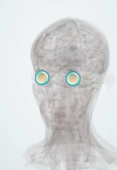
See it in 3D
Who gets it
Increasing worldwide with the rise of myopia; especially common in East Asia.
Causes
- Excessive axial elongation of the eye
- Genetic predisposition
- Environmental factors in childhood (near work, little time outdoors)
Risk factors
- High myopia in childhood
- Myopic parents
- Rapid progression
Pathophysiology
Stretching of the eye wall thins the sclera, choroid and retina; Bruch membrane cracks (lacquer cracks) and new vessels can grow through them. The vitreous and retina are pulled at the posterior pole and periphery, causing schisis, holes and detachment.
Symptoms and signs
- Very poor uncorrected vision
- Gradual loss of central vision
- Sudden distortion or a central spot (myopic new vessels)
- Floaters and flashes (detachment risk)
Diagnosis
- Clinical
- Tilted disc with peripapillary atrophy, posterior staphyloma, lacquer cracks, patchy chorioretinal atrophy and a dark macular scar (Fuchs spot).
- Tests
- Refraction and axial length measurement; visual fields.
- Imaging
- OCT for myopic traction and new vessels; angiography when new vessels are suspected.
Differential diagnosis
- Age-related macular degeneration
- Other causes of choroidal neovascularisation
- Inherited retinal dystrophies
New vessels at the macula: myopic or age-related?
| Feature | Myopic choroidal new vessels | Neovascular age-related macular degeneration |
|---|---|---|
| Typical age | Often under 50 | Usually over 60 |
| Refraction | High myopia with a long eye | Usually no high myopia |
| Background fundus | Lacquer cracks, thin choroid, patchy atrophy | Drusen and pigment changes |
| Lesion | Small, flat, near a lacquer crack; may leave a dark Fuchs spot | Larger, with more exudate and haemorrhage |
| Response to anti-VEGF | Usually good with few injections | Often needs long-term injections |
Goal of treatment
Correct the refraction, detect and treat complications early and slow progression in children.
Treatment
- Conservative
- Spectacles or contact lenses; low vision aids when needed.
- Medical
- Intravitreal anti-VEGF injections for myopic choroidal new vessels.
- Surgical
- Vitrectomy for myopic traction or macular hole; repair of retinal detachment.
- Lifestyle
- Myopia control measures in childhood (outdoor time, specialist optical or drug treatment).
Complications
- Retinal detachment
- Myopic choroidal neovascularisation
- Macular atrophy
- Glaucoma
- Early cataract
Common traps
- Glasses solve pathological myopia: they correct focus, but the eye keeps stretching and the degeneration of choroid and retina continues, so regular examination for complications is needed
- Sudden distortion in a high myope is age-related macular degeneration: in younger high myopes it is usually myopic choroidal new vessels through a lacquer crack, treated promptly with anti-VEGF
- Glaucoma is easy to judge from the disc in high myopia: a tilted disc with peripapillary atrophy hides cupping, and myopic retinal changes cause field defects of their own, so OCT and fields must be read with caution
- Flashes and floaters in a high myope are harmless vitreous floaters: thin peripheral retina raises the risk of a rhegmatogenous detachment, so a dilated retinal examination is needed
- Myopia progression in children cannot be changed: more time outdoors and specialist optical or drug treatments slow progression, which lowers the later risk of complications
Prognosis
Variable; complications can cause severe central visual loss, but many respond to timely treatment.
Test yourself
Think of the answer, then tap the question to see it.
Why does an eye that keeps lengthening develop retinal and choroidal damage?
The sclera keeps stretching, but the retina, choroid and Bruch membrane cannot stretch as much, so they thin, crack and tear.
A 38-year-old high myope has sudden distortion in one eye. Most likely cause and next step?
Myopic choroidal new vessels through a lacquer crack; OCT and angiography, then prompt intravitreal anti-VEGF.
Why is glaucoma hard to detect in pathological myopia?
Tilted discs with peripapillary atrophy mask cupping, and myopic retinal changes cause their own field defects, so both main signs mislead.
A high myope reports new flashes and a shower of floaters. What next?
Same-day dilated retinal examination for a tear or rhegmatogenous detachment, which thin peripheral retina makes more likely.
Which childhood measures help slow myopia progression?
More time outdoors, plus specialist optical or drug myopia-control treatment when progression is rapid.
Connections
In physiology1
Educational information, not medical advice. For any symptom, see a health professional.
Sources: Degenerative Myopia (StatPearls Publishing (NCBI Bookshelf), 2022).
Spotted a mistake?We are always improving for the community. Tell us what is wrong and our team will review it.Report a mistake
Representative model, for educational purposes only.