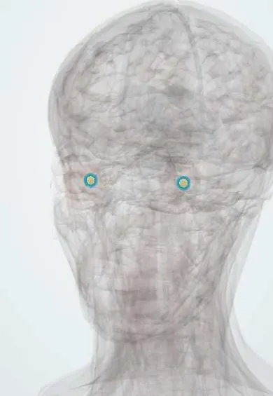
Macula !
The macula is a small yellowish spot in the centre of the retina, with a central pit (the fovea); it gives the sharp central vision used to read and see faces.
Opens in the Durgical app, no sign-up needed.
What it is
A 5 mm yellowish spot in the centre of the retina, with a central pit (the fovea).
What it does
Gives sharp central and colour vision for reading and faces.
Key facts
- Where
- Posterior pole, ~4 mm temporal to the optic disc; fovea at its centre
- Blood supply
- Choroid (fovea has no retinal vessels)
- Function
- Sharp central and colour vision (cones only at the fovea)
Relations
- Superficial
- Retinal pigment epithelium, Bruch membrane and the choriocapillaris, which supplies the fovea
- Deep
- Vitreous body; abnormal adhesion of the posterior hyaloid can pull on it (vitreomacular traction, macular hole)
- Medial
- Optic disc, on the nasal side, joined to it by the papillomacular bundle of nerve fibres
- Lateral
- Temporal retina, whose nerve fibres arch above and below the macula and meet at the horizontal raphe
How to find it
On fundoscopy it is the darker zone temporal to the disc, free of large vessels, with a bright foveal reflex at its centre in young eyes. Asking the patient to look straight at the ophthalmoscope light brings it into view.
Examination
- Amsler grid: wavy or missing lines (metamorphopsia, scotoma) suggest macular disease
- Visual acuity and colour vision: both drop early in macular disease
- Photostress recovery: slow return of acuity after a bright light points to the macula rather than the optic nerve
On imaging
OCT shows the foveal pit, fluid, drusen, holes and membranes in cross-section; fluorescein angiography shows leaking new vessels; fundus autofluorescence maps atrophy of the pigment epithelium.
Common problems
- Age-related macular degeneration: central vision blurs or distorts (straight lines look wavy); the wet form can worsen fast.
- Diabetic macular oedema: fluid swells the macula and blurs reading vision.
- Macular hole or pucker: a gap or wrinkle in the central retina.
How it is diagnosed
A grid test (Amsler) at home, dilated eye exam and OCT scans.
Treatment
- Regular injections into the eye (anti-VEGF) for wet macular degeneration and macular oedema.
- Vitamin supplements in some dry macular degeneration.
- Vitrectomy surgery for holes and puckers.
Clinical notes
- Hydroxychloroquine and chloroquine can damage it (bull's-eye maculopathy), so long-term users are screened
- Macular star: exudates radiating around the fovea in severe hypertensive retinopathy and in neuroretinitis
- Central serous chorioretinopathy: fluid under the macula, typically in young men and linked to steroid use
- Cystoid macular oedema can follow cataract surgery or uveitis
Anatomical variants
- A cilioretinal artery, from the ciliary (choroidal) circulation and entering at the disc edge, supplies part of the macula in a sizeable minority of eyes (figures vary by method); it can save central vision in a central retinal artery occlusion
Development
The fovea is immature at birth; its cones pack closer and the pit deepens over the first years of life.
Related conditions
- Tay–Sachs disease
- Sandhoff disease
- GM2 activator deficiency (GM2 gangliosidosis, AB variant)
- GM1 gangliosidosis
- Diabetic retinopathy
- Age-related macular degeneration
Test yourself
Think of the answer, then tap the question to see it.
Why does a cherry-red spot appear at the macula in central retinal artery occlusion?
The inner retina turns pale, and the thin fovea lets the red choroid show through.
How do the dry and wet forms of age-related macular degeneration differ?
Dry: drusen and slow atrophy; wet: new choroidal vessels grow through Bruch membrane and leak or bleed, treated with anti-VEGF injections.
Which simple test detects distortion from macular disease?
The Amsler grid: wavy or missing lines (metamorphopsia, scotoma) suggest macular disease.
Why are long-term users of hydroxychloroquine screened at the macula?
The drug can damage the macula, giving a bull's-eye maculopathy.
Educational information, not medical advice. For any symptom, see a health professional.
Its names
- Official Latin (TA2)
- macula lutea TA2 6784
- Official Spanish (DeCS)
- Mácula Lútea DeCS D008266
- English (TA2)
- Macula
- Also called
- macula lutea · yellow spot
Related
Sources: Terminologia Anatomica (TA2); DeCS; Anatomy, Head and Neck, Eye Fovea; Macular Degeneration.
Spotted a mistake?We are always improving for the community. Tell us what is wrong and our team will review it.Report a mistake
Representative model, for educational purposes only.