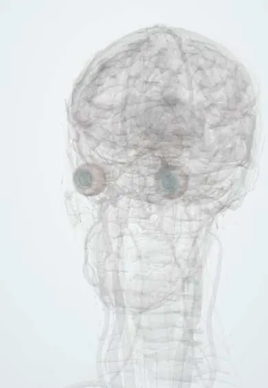
Anterior chamber !
The anterior chamber is the fluid-filled space between the cornea and the iris; its fluid drains at the edge, and poor drainage raises eye pressure (glaucoma).
Official name: Anterior chamber of eyeball
Opens in the Durgical app, no sign-up needed.
What it is
The fluid-filled space between the cornea and the iris; its drainage angle is where glaucoma starts.
What it does
The fluid drains at its edge (the angle); if drainage fails, pressure rises (glaucoma).
Key facts
- Where
- Between the cornea and the iris
- Function
- Holds aqueous humour; drains at the iridocorneal angle
Relations
- In front
- Back surface of the cornea (endothelium), which forms its front wall
- Behind
- Front of the iris and, within the pupil, the front of the lens; behind the iris lies the posterior chamber, joined to it through the pupil
- Contents
- Aqueous humour, flowing from the pupil to the drainage angle at its outer edge, where the trabecular meshwork and Schlemm canal take it out of the eye
How to find it
Seen through the clear cornea as the space in front of the iris. Its depth is judged at the slit lamp by comparing the gap between cornea and iris at the limbus with corneal thickness (van Herick method), or with a penlight shone from the temporal side: in a shallow chamber the forward-bowed iris casts a shadow on its nasal half.
Examination
- Oblique penlight (shadow) test: a shadow over the nasal iris suggests a shallow chamber and a narrow angle
- Slit-lamp exam: cells and flare in the light beam point to anterior uveitis; a level of blood (hyphema) or pus (hypopyon) settles at the bottom with the patient upright
On imaging
Anterior segment OCT and ultrasound biomicroscopy measure its depth and show the angle; B-scan ultrasound checks the back of the eye when a hyphema blocks the view.
Common problems
- Open-angle glaucoma: the meshwork drains poorly, pressure creeps up and side vision fades with no pain.
- Angle-closure glaucoma: the iris blocks the angle; sudden eye pain, redness, halos, headache and vomiting: an emergency.
How it is diagnosed
Eye-pressure measurement (tonometry), viewing the angle with a special lens (gonioscopy), optic disc exam and visual fields.
Treatment
- Daily eye drops to lower the pressure.
- Laser to open the drainage (trabeculoplasty) or make a hole in the iris (iridotomy).
- Surgery to make a new drainage path.
Clinical notes
- Traumatic hyphema: the blood can clog the meshwork and raise eye pressure, and rebleeding in the first days is a classic complication
- In sickle cell disease or trait, sickled red cells drain poorly, so even a small hyphema can raise the pressure sharply
- Dilating the pupil in an eye with a shallow chamber can trigger acute angle-closure glaucoma
- Paracentesis, a fine needle entry at the limbus, samples the aqueous or lowers the pressure quickly
Development
Opens as a cleft in the mesenchyme between the developing cornea and the lens; faulty migration of neural crest cells here causes anterior segment dysgenesis (Axenfeld–Rieger, Peters anomaly), which often leads to glaucoma.
Related conditions
Test yourself
Think of the answer, then tap the question to see it.
What is the route of aqueous humour through the anterior chamber?
Made by the ciliary body, it fills the posterior chamber, passes through the pupil into the anterior chamber and leaves at the drainage angle.
What do a hyphema and a hypopyon in the anterior chamber consist of?
A hyphema is layered blood and a hypopyon is layered pus at the bottom of the anterior chamber.
What can dilating the pupil trigger in an eye with a shallow anterior chamber?
Acute angle-closure glaucoma, because a shallow chamber means a narrow angle.
Why can even a small hyphema raise eye pressure sharply in sickle cell disease or trait?
Sickled red cells drain poorly through the trabecular meshwork.
Educational information, not medical advice. For any symptom, see a health professional.
Its names
- Official Latin (TA2)
- camera anterior bulbi oculi TA2 6792
- Official Spanish (DeCS)
- Cámara Anterior DeCS D000867
- English (TA2)
- Anterior chamber of eyeball
- Also called
- camera oculi anterior
Related
Sources: Terminologia Anatomica (TA2); DeCS; Hyphema; Physiology, Aqueous Humor Circulation.
Spotted a mistake?We are always improving for the community. Tell us what is wrong and our team will review it.Report a mistake
Representative model, for educational purposes only.