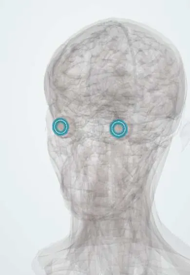
Drainage angle !
The drainage angle is the corner where the iris meets the cornea; it drains the eye's fluid, and when it clogs or closes, eye pressure rises (glaucoma).
Official name: Iridocorneal angle
Opens in the Durgical app, no sign-up needed.
What it is
The corner where the iris meets the cornea, with a sponge-like filter and a ring canal.
What it does
Drains the aqueous humour; when it clogs or closes, eye pressure rises (glaucoma).
Key facts
- Where
- Junction of the cornea and iris (limbus)
- Function
- Trabecular meshwork -> Schlemm's canal -> aqueous veins; blockage raises eye pressure (glaucoma)
Relations
- Course
- Aqueous crosses the uveal, corneoscleral and juxtacanalicular layers of the trabecular meshwork into Schlemm canal; collector channels and aqueous veins then carry it to the episcleral veins
- In front
- Schwalbe line, where Descemet membrane ends at the edge of the cornea
- Behind
- Iris root and the ciliary body band
- Superficial
- Sclera at the limbus, inside which Schlemm canal runs as a ring
- Deep
- Anterior chamber, whose aqueous enters the meshwork
How to find it
It cannot be seen straight on, because its light is totally reflected inside the cornea; a gonioscopy lens with mirrors placed on the eye shows it at the slit lamp. On the outside it lies under the limbus, the grey-blue junction of cornea and sclera.
Examination
- Gonioscopy: a visible scleral spur or meshwork means an open angle; only Schwalbe line, or no structures, means a narrow or closed angle; pressing on the cornea (indentation) tells reversible from scarred closure
- Van Herick estimate at the slit lamp: a peripheral chamber depth under a quarter of corneal thickness suggests a narrow angle
- Tonometry: raised pressure supports poor outflow, although glaucoma can occur at normal pressures
On imaging
Anterior segment OCT (no contact) and ultrasound biomicroscopy measure the angle width and show causes of closure such as plateau iris; they complement gonioscopy rather than replace it.
Common problems
- Open-angle glaucoma: the meshwork drains poorly, pressure creeps up and side vision fades with no pain.
- Angle-closure glaucoma: the iris blocks the angle; sudden eye pain, redness, halos, headache and vomiting: an emergency.
How it is diagnosed
Eye-pressure measurement (tonometry), viewing the angle with a special lens (gonioscopy), optic disc exam and visual fields.
Treatment
- Daily eye drops to lower the pressure.
- Laser to open the drainage (trabeculoplasty) or make a hole in the iris (iridotomy).
- Surgery to make a new drainage path.
Clinical notes
- Laser peripheral iridotomy makes a small hole in the iris so aqueous bypasses the pupil and the iris falls back; the other eye is usually treated as well
- Laser trabeculoplasty, trabeculectomy (a filtering bleb under the conjunctiva) and tube shunts all aim to improve outflow
- Secondary glaucoma: pigment, exfoliation material, blood, inflammatory cells or lens protein can clog the meshwork; new vessels in diabetes or vein occlusion can seal the angle (neovascular glaucoma)
- Angle recession after blunt trauma, often with a hyphema, can cause glaucoma years later
- Drugs that dilate the pupil (anticholinergic or sympathomimetic) can set off angle closure in an eye with a narrow angle
Anatomical variants
- Iris processes: fine strands from the iris root to the scleral spur or meshwork, common on gonioscopy and normal unless broad and numerous
- Posterior embryotoxon: a thickened Schwalbe line set forward, seen as a white ring just inside the limbus; present in a minority of normal eyes, and part of Axenfeld–Rieger and Alagille syndromes
Development
Forms from neural crest mesenchyme; incomplete maturation of the meshwork causes primary congenital glaucoma (large hazy cornea, tearing, light sensitivity), and anterior segment dysgenesis (Axenfeld–Rieger) carries a glaucoma risk.
Related conditions
Test yourself
Think of the answer, then tap the question to see it.
Which structures are seen on gonioscopy of the drainage angle, from the iris outwards?
Iris root, ciliary body band, scleral spur, trabecular meshwork (the pigmented band) and Schwalbe line.
What is the conventional outflow route of aqueous humour from the drainage angle?
Trabecular meshwork, Schlemm canal, then aqueous and episcleral veins; a smaller share leaves through the ciliary muscle (uveoscleral route).
What are the features of acute angle closure?
Red painful eye, hazy cornea, mid-dilated poorly reacting pupil, rock-hard globe, headache and vomiting: a sight-threatening emergency.
How does open-angle glaucoma differ from angle closure at the drainage angle?
The angle looks normal but drains poorly; the disease is silent until peripheral vision is lost.
What can cause glaucoma years after blunt trauma with a hyphema?
Angle recession: damage to the drainage angle at the time of injury.
Educational information, not medical advice. For any symptom, see a health professional.
Its names
- Official Latin (TA2)
- angulus iridocornealis TA2 6793
- Official Spanish (DeCS)
- Ángulo iridocorneal DeCS D014129
- English (TA2)
- Iridocorneal angle
- Also called
- trabecular meshwork · Schlemm's canal · angulus iridis
Related
Sources: Terminologia Anatomica (TA2); DeCS; Open Angle Glaucoma; Acute Angle-Closure Glaucoma; Physiology, Aqueous Humor Circulation.
Spotted a mistake?We are always improving for the community. Tell us what is wrong and our team will review it.Report a mistake
Representative model, for educational purposes only.