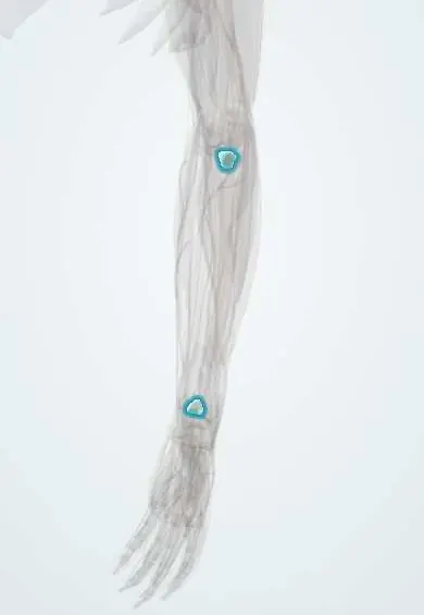
Radial head cartilage !
The radial head cartilage covers the top of the radius at the elbow; it lets the radius spin against the humerus and ulna as the forearm turns.
Official name: Cartilage of the radial head
Opens in the Durgical app, no sign-up needed.
What it is
The smooth cap on the disc-shaped top of the radius.
What it does
Lets the radius spin against the humerus and ulna when the forearm turns.
Key facts
- Where
- Concave top and rim of the radial head
- Function
- Glides on the capitulum and turns in the radial notch and annular ligament
Relations
- Above
- Capitulum of the humerus, across the radiocapitellar joint
- Medial
- Radial notch of the ulna, forming the proximal radioulnar joint
- Lateral
- Annular ligament encircling the rim, with the RCL (elbow) and the supinator over it
- Below
- Radial neck, where the posterior interosseous nerve winds round to enter the supinator
- Behind
- Anconeus and the LUCL (lateral ulnar collateral ligament)
How to find it
It lies in the lateral 'soft spot', the triangle between the lateral epicondyle, the radial head and the tip of the olecranon. A thumb placed just below the lateral epicondyle feels the head turn as the forearm is pronated and supinated; the same spot is used for elbow aspiration and an arthroscopy portal.
On imaging
AP and lateral X-rays, plus a radiocapitellar (Greenspan) oblique view that projects the head clear of the coronoid. After trauma, a lifted anterior fat pad ('sail') or any visible posterior fat pad means an effusion and, in an adult, a likely hidden radial head fracture.
Clinical notes
- Essex-Lopresti injury: a radial head fracture with tearing of the interosseous membrane and the distal radioulnar joint; removing the head then lets the radius migrate upward, so it is fixed or replaced rather than excised
- The posterior interosseous nerve crosses the radial neck: lateral approaches are made with the forearm pronated, which moves the nerve forward and away
- In the terrible triad (posterior elbow dislocation with radial head and coronoid fractures) the radial head is fixed or replaced to restore stability
Development
Its ossification centre appears in early childhood (the 'R' of CRITOE); children tend to fracture the radial neck rather than the head, and the physis below the head can be mistaken for a fracture.
Related conditions
Test yourself
Think of the answer, then tap the question to see it.
What are the most common elbow fractures in adults, and how are they graded?
Radial head fractures, graded by Mason: I undisplaced, II displaced partial-head, III comminuted, IV with a dislocation.
Which nerve crosses the radial neck, and how is it protected in lateral approaches?
The posterior interosseous nerve; the forearm is pronated, which moves the nerve forward and away.
After elbow trauma in an adult, what does a visible posterior fat pad suggest?
An effusion and a likely hidden radial head fracture.
Educational information, not medical advice. For any symptom, see a health professional.
Its names
- Official Latin (TA2)
- cartilago articularis capitis radii TA2 382
- Official Spanish (DeCS)
- Cartílago articular de la cabeza del radio
- English (TA2)
- Cartilage of the radial head
Related
Sources: Terminologia Anatomica (TA2); Anatomy, Shoulder and Upper Limb, Elbow Joint.
Spotted a mistake?We are always improving for the community. Tell us what is wrong and our team will review it.Report a mistake
Representative model, for educational purposes only.