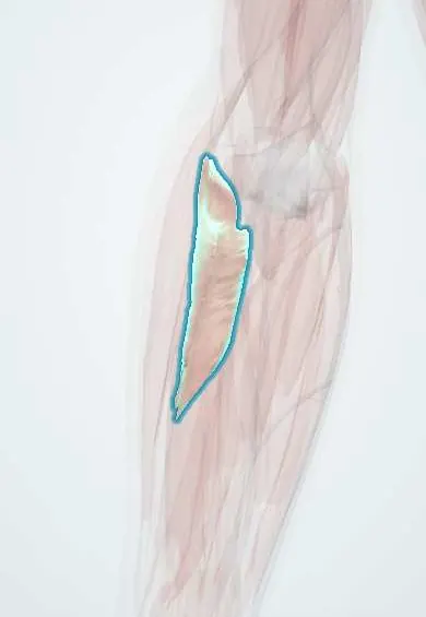
Supinator !
The supinator wraps around the top of the radius below the elbow; it turns the palm up, and a branch of the radial nerve passes through it.
Opens in the Durgical app, no sign-up needed.
What it is
A short muscle wrapped around the top of the radius; the posterior interosseous nerve passes through it.
What it does
Turns the palm up.
Key facts
- Origin
- Lateral epicondyle, radial collateral and annular ligaments, supinator crest of the ulna
- Insertion
- Proximal third of the radius (lateral, posterior and anterior)
- Action
- Supinates the forearm
- Nerve
- Deep branch of the radial nerve / posterior interosseous nerve (C5–C6)
- Blood supply
- Radial recurrent artery, recurrent interosseous artery
Relations
- Contents
- The deep branch of the radial nerve (posterior interosseous nerve), running between its superficial and deep layers
- Superficial
- Brachioradialis, ECRL, ECRB and extensor digitorum; in front, the radial artery and the superficial radial nerve lie on it
- Deep
- Neck and upper shaft of the radius, the annular ligament and the elbow capsule
- Below
- At its lower border the posterior interosseous nerve emerges among APL, EPB and the finger extensors, joined by the posterior interosseous artery
How to find it
Not felt as a separate belly; it wraps the upper third of the radius under the mobile wad. In the posterolateral (Thompson) approach it is exposed between ECRB and extensor digitorum, with the posterior interosseous nerve seen running through it.
Examination
- Resisted supination with the elbow straight: pain in the upper outer forearm suggests radial tunnel syndrome
- Point tenderness over the supinator, a few finger-widths below the lateral epicondyle, favours radial tunnel syndrome over tennis elbow
On imaging
Rarely assessed for its own sake: imaging looks at the posterior interosseous nerve entering it under the arcade of Frohse. Ultrasound follows the nerve through the muscle and can show it swollen just before the arcade, or a compressing ganglion or lipoma. On MRI, denervation of the extensors beyond it shows as oedema, later as fatty atrophy.
Clinical notes
- PIN syndrome: weak finger and thumb extension (finger drop), wrist extension drifting radially, no sensory loss
- Radial tunnel syndrome: pain without weakness; compression sites are fibrous bands at the radiocapitellar joint, the recurrent radial vessels (leash of Henry), the ECRB edge, the arcade of Frohse and the lower edge of the supinator
- Monteggia fracture: the dislocated radial head can stretch the posterior interosseous nerve as it runs through the muscle
Anatomical variants
- The arcade of Frohse varies from thin and membranous to thick and tendinous, which may explain why compression affects some people only
Related conditions
Test yourself
Think of the answer, then tap the question to see it.
What passes between the two layers of the supinator?
The deep branch of the radial nerve (posterior interosseous nerve).
What is the arcade of Frohse?
The fibrous upper edge of the superficial layer of the supinator, the most common compression site of the posterior interosseous nerve.
How does radial tunnel syndrome differ from posterior interosseous nerve syndrome?
Radial tunnel syndrome is pain without weakness; PIN syndrome gives weak finger and thumb extension without sensory loss.
When is the supinator the main supinator, and when does the biceps take over?
In slow, unresisted supination with the elbow straight; the biceps takes over with the elbow flexed or against resistance.
Educational information, not medical advice. For any symptom, see a health professional.
Its names
- Official Latin (TA2)
- supinator TA2 2512
- Official Spanish (DeCS)
- Músculo supinador
- English (TA2)
- Supinator
Related
Sources: Terminologia Anatomica (TA2); Anatomy, Shoulder and Upper Limb, Forearm Muscles; Anatomy, Shoulder and Upper Limb, Radial Nerve.
Spotted a mistake?We are always improving for the community. Tell us what is wrong and our team will review it.Report a mistake
Representative model, for educational purposes only.