Joints and ligaments · Upper limb
Annular ligament !
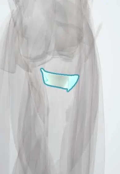
The annular ligament is a ring around the top of the radius at the elbow; it holds the radial head against the ulna while it spins.
Official name: Annular ligament of the radius
Opens in the Durgical app, no sign-up needed.
What it is
A ring of ligament around the head of the radius, fixed to the ulna at both ends.
What it does
Holds the radial head against the ulna while it spins.
Key facts
- Attaches
- Anterior and posterior margins of the radial notch of the ulna, encircling the radial head
- Function
- Holds the radial head against the ulna while it spins
- Tight in
- Constant during rotation
Relations
- Contents
- Radial head rim, which turns within it; its smooth inner surface continues the radial notch
- Medial
- Radial notch of the ulna, to whose front and back edges it attaches
- Above
- RCL (elbow) blending into it from the lateral epicondyle; the capitulum above the radial head
- Below
- Radial neck, the quadrate ligament and the synovial pouch that bulges below it (sacciform recess)
- Lateral
- Supinator, which partly arises from it, and the common extensor origin
- In front
- Distal biceps tendon heading for the radial tuberosity; the posterior interosseous nerve passes round the radial neck to enter the supinator under the arcade of Frohse
- Behind
- Anconeus and the LUCL
How to find it
Place a finger in the small hollow just below the lateral epicondyle at the back of the extended elbow and turn the forearm: the radial head rolls under the finger inside this ligament. At surgery it lies deep to the supinator and the common extensor origin.
Examination
- Reduction of a pulled elbow confirms the diagnosis: supination with flexion, or hyperpronation, of the forearm; a click over the radial head and the child using the arm again within minutes are typical (studies suggest hyperpronation succeeds slightly more often)
On imaging
On every elbow X-ray the radiocapitellar line, drawn along the radial neck, must pass through the capitulum in all views; if not, the radial head is dislocated and a Monteggia ulna fracture is sought. In a typical pulled elbow X-rays are normal; ultrasound can show the ligament caught in the joint.
Common problems
- Nursemaid's elbow: in young children a sudden pull on the hand slips the radial head partly out of the ring.
- Torn with radial head dislocations and some forearm fractures.
How it is diagnosed
A child who will not use the arm after a pull, with normal-looking X-rays. MRI for adult injuries.
Treatment
- A gentle reduction move by a clinician usually fixes nursemaid's elbow at once.
- Repair or reconstruction in complex adult injuries.
Clinical notes
- Bado classification of Monteggia injuries: I anterior radial head dislocation (most common in children), II posterior, III lateral, IV with fractures of both bones
- A missed, chronic Monteggia lesion may need annular ligament reconstruction (for example with a strip of triceps fascia) together with correction of the ulna
- Congenital radial head dislocation (usually posterior, often both sides, with a dome-shaped head) differs from a traumatic one
Anatomical variants
- Its shape varies: dissection and MRI studies describe single, two-lobed and other forms
Memory aid
MUGR: Monteggia = Ulna fracture (with radial head dislocation), Galeazzi = Radius fracture (with distal radioulnar joint injury).
Related conditions
Test yourself
Think of the answer, then tap the question to see it.
What does the annular ligament attach to, and what does it do?
The front and back margins of the radial notch of the ulna, encircling the radial head; it holds the radial head against the ulna while it spins.
What happens to the annular ligament in a pulled elbow (radial head subluxation)?
In children of about 1 to 4 years, a pull on the pronated hand slips the annular ligament up over the radial head into the joint.
How is a pulled elbow reduced, confirming the diagnosis?
By supination with flexion, or hyperpronation, of the forearm; a click over the radial head and the child using the arm again confirm it.
Which fracture-dislocation tears or displaces the annular ligament?
The Monteggia injury: an ulna fracture with radial head dislocation; type I (anterior) is the most common in children.
Educational information, not medical advice. For any symptom, see a health professional.
Its names
- Official Latin (TA2)
- ligamentum anulare radii TA2 1778
- Official Spanish (DeCS)
- Ligamento anular del radio
- English (TA2)
- Annular ligament of the radius
Related
Sources: Terminologia Anatomica (TA2); Anatomy, Shoulder and Upper Limb, Elbow Annular Ligament; Anatomy, Shoulder and Upper Limb, Proximal Radio-Ulnar Joint; Anatomy of the Human Body (20th ed.): Surface Anatomy of the Upper Extremity (Lea & Febiger (Gray's Anatomy, 1918), 1918) · adapted, Public domain; 9.6 Anatomy of Selected Synovial Joints (OpenStax (Rice University), 2022) · adapted, CC BY 4.0.
Spotted a mistake?We are always improving for the community. Tell us what is wrong and our team will review it.Report a mistake
Representative model, for educational purposes only.