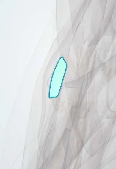
Joints and ligaments · Upper limb
RCL (elbow) !
The radial collateral ligament is on the outer side of the elbow; it stops the joint opening outward and blends into the ring around the radius.
Official name: Radial (lateral) collateral ligament of the elbow
Opens in the Durgical app, no sign-up needed.
What it is
A band on the outer side of the elbow, from the lateral epicondyle into the annular ligament.
What it does
Stops the elbow opening on the outer side.
Key facts
- Attaches
- Lateral epicondyle to the annular ligament (some texts: anterior, middle and posterior bundles reaching the ulna around the radial notch and the olecranon)
- Function
- Resists varus stress
- Tight in
- Throughout the arc (nearly isometric)
Relations
- Course
- From the lateral epicondyle, beneath the common extensor origin, down into the annular ligament around the radial head
- Superficial
- Common extensor tendon, especially extensor carpi radialis brevis (ECRB), whose fibres blend with it; the supinator arises partly from it
- Deep
- Lateral capsule and the radiocapitellar joint (radial head cartilage on the capitulum)
- Behind
- LUCL and the anconeus
How to find it
Find the lateral epicondyle, then the radial head in the hollow just below it while the forearm is turned: the ligament spans these two points and fans into the annular ligament. At surgery it lies deep to the common extensor origin, exposed in the interval between anconeus and ECU (Kocher).
Examination
- Varus stress test: elbow at 20–30° of flexion with the humerus fully internally rotated; lateral opening compared with the other side suggests a lateral collateral ligament injury
On imaging
Coronal MRI shows the RCL and LUCL together: thickening and signal change in chronic lateral epicondylitis, or detachment from the lateral epicondyle (the usual site of failure) after a dislocation; stress views show lateral opening.
Common problems
- Torn in elbow dislocations, usually together with the LUCL.
- Damage can leave the elbow unstable when twisting with the arm straight (posterolateral rotatory instability).
How it is diagnosed
Pivot-shift or chair push-up tests; MRI shows the ligament complex.
Treatment
- Brace and guided motion after most simple dislocations.
- Repair or reconstruction with a tendon graft for ongoing instability.
Clinical notes
- Lateral approaches to the radial head open the capsule in front of the LUCL, above the equator of the radial head with the forearm pronated, to avoid causing instability
- Overly deep debridement in surgery for lateral epicondylitis (tennis elbow) can injure the lateral ligament complex and cause instability
- In elbow dislocations the lateral complex usually fails first, at its humeral origin; in the terrible triad it is reattached with anchors or bone tunnels
Test yourself
Think of the answer, then tap the question to see it.
Where does the radial collateral ligament of the elbow insert distally, and why does it matter?
Into the annular ligament, not the radius, so the radius stays free to rotate.
What stress does the radial collateral ligament of the elbow resist?
Varus stress; it is nearly isometric, taut throughout the arc of motion.
Where does the radial collateral ligament of the elbow arise, and why do reconstructions use that point?
At the isometric point of the lateral epicondyle, the centre of the capitulum's arc; the humeral tunnel is placed there.
Educational information, not medical advice. For any symptom, see a health professional.
Its names
- Official Latin (TA2)
- ligamentum collaterale radiale cubiti TA2 1777
- Official Spanish (DeCS)
- Ligamento colateral radial del codo
- English (TA2)
- Radial (lateral) collateral ligament of the elbow
- Also called
- lateral collateral ligament of elbow · RCL · radial collateral ligament of elbow · lateral collateral ligament complex
Related
Sources: Terminologia Anatomica (TA2); Anatomy, Shoulder and Upper Limb, Elbow Collateral Ligaments; 9.6 Anatomy of Selected Synovial Joints (OpenStax (Rice University), 2022) · adapted, CC BY 4.0; Anatomy of the Human Body (20th ed.): Surface Anatomy of the Upper Extremity (Lea & Febiger (Gray's Anatomy, 1918), 1918) · adapted, Public domain.
Spotted a mistake?We are always improving for the community. Tell us what is wrong and our team will review it.Report a mistake
Representative model, for educational purposes only.