Cardiovascular system · Head and neck
Central retinal artery !
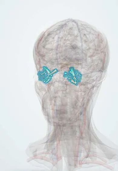
The central retinal artery runs inside the optic nerve and spreads over the retina to feed it; a blockage causes sudden, painless loss of vision.
Opens in the Durgical app, no sign-up needed.
What it is
Vessels running inside the optic nerve and spreading over the retina.
What it does
Feed the inner retina; a blocked artery causes sudden painless vision loss.
Key facts
- Origin
- Ophthalmic artery; enters the optic nerve ~1 cm behind the eye
- Branches
- Four branches over the retina (superior/inferior, nasal/temporal); end arteries
- Supplies
- Inner retina (occlusion = sudden painless loss of vision; cherry-red spot)
Relations
- Course
- Arises from the ophthalmic artery near the optic canal, runs forward below the optic nerve, pierces its dural sheath about 1 cm behind the eyeball and travels in the centre of the nerve to emerge at the optic disc, where it divides
- Deep
- Over the retina its branches run in the nerve fibre layer under the internal limiting membrane, facing the vitreous body; where they cross veins they share a sheath (arteriovenous crossings)
How to find it
On fundoscopy its branches are the narrower, brighter red vessels leaving the disc, with a central light reflex; they are thinner than the veins beside them. Following the arcades out from the disc shows the four main branches.
Examination
- Fundoscopy: whitened, swollen retina, cherry-red spot, thin arteries, sometimes an embolus visible at a branch point
- Swinging flashlight test: a relative afferent pupillary defect in the affected eye
- Inflammatory markers and temporal artery assessment in older patients, looking for giant cell arteritis
On imaging
Fluorescein angiography shows delayed arterial filling; OCT shows a thick, bright inner retina early and thinning later; the stroke workup includes carotid imaging, an ECG and imaging of the heart.
Clinical notes
- Branch retinal artery occlusion causes a sector of field loss matching the territory supplied
- Hypertensive and arteriosclerotic changes are seen directly in its branches: narrowing, copper or silver wiring, and nicking where arteries cross veins
- Central retinal vein occlusion, of its companion vein, looks very different: swollen tortuous veins and haemorrhages across the whole retina
Anatomical variants
- Cilioretinal artery: a branch of the posterior ciliary circulation emerging at the temporal edge of the disc in a sizeable minority of eyes (figures vary between texts); it can spare central vision in an occlusion
Development
It is the persistent proximal part of the hyaloid artery, whose segment crossing the vitreous to the lens regresses before birth.
Related conditions
- Central retinal artery occlusion
- Hypertensive retinopathy
- Transient ischaemic attack
- Diabetic retinopathy
- Giant cell arteritis
Test yourself
Think of the answer, then tap the question to see it.
What are the signs of central retinal artery occlusion?
Sudden painless loss of vision in one eye, a pale retina with a cherry-red spot, a relative afferent pupillary defect and segmented (boxcar) blood columns.
Which cause must be ruled out in an older patient with central retinal artery occlusion?
Giant cell arteritis; other main causes are emboli from the carotid artery (Hollenhorst plaques) or the heart.
What is amaurosis fugax and how is it investigated?
A brief curtain over one eye: a transient ischaemic attack of the retina, with the same workup as a stroke.
How can a cilioretinal artery change the effect of a central retinal artery occlusion?
This branch of the posterior ciliary circulation, present in a sizeable minority of eyes, can spare central vision.
Educational information, not medical advice. For any symptom, see a health professional.
Its names
- Official Latin (TA2)
- arteria centralis retinae TA2 4470
- Official Spanish (DeCS)
- Arteria Retiniana DeCS D012161
- English (TA2)
- Central retinal artery
- Also called
- Zinn's artery
Related
Sources: Terminologia Anatomica (TA2); DeCS; Central Retinal Artery Occlusion; Anatomy, Head and Neck, Eye Arteries; Tratado de anatomía humana (9.ª ed.), tomo II, Libro IV, sección 2 Arterias, caps. I-III (sistema de la arteria pulmonar; aorta y ramas del cayado hasta la carótida interna).
Spotted a mistake?We are always improving for the community. Tell us what is wrong and our team will review it.Report a mistake
Representative model, for educational purposes only.