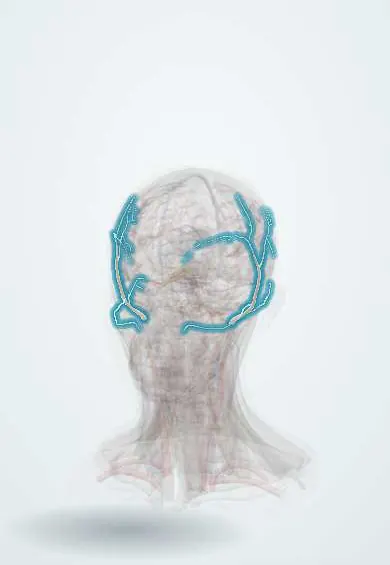
Giant cell arteritis !
Giant cell arteritis is inflammation of large arteries, such as the one at the temple, in people over 50; it causes headache and can cause sudden blindness.
Updated 2026-10-29
Structures involved
Tap one to see what it is and open it in 3D.
- Superficial temporal artery
- Ophthalmic artery
- Optic nerve
- Central retinal artery
- Optic disc
- Maxillary artery
- Masseter
- External carotid artery
- Subclavian artery
In physiology
Why it happens
- Granulomatous inflammation: caseating vs non-caseating, the causes to know
- Systemic effects of inflammation: fever, acute-phase proteins, leukocytosis, sepsis
- C-reactive protein (CRP) as a lab test
- Blood vessels: artery, vein and capillary layers
What it is
Granulomatous vasculitis of large and medium arteries in people over 50, typically the branches of the external carotid artery such as the superficial temporal artery. Its feared complication is sudden permanent blindness.
Who gets it
The commonest systemic vasculitis in older adults in Europe and North America, more frequent in women and in people of Northern European descent. It is reported less often in Latin America but occurs and may be underdiagnosed.
Causes
- Unknown; immune-mediated granulomatous inflammation in a genetically susceptible older person
- Strong association with polymyalgia rheumatica
Risk factors
- Age over 50 (peak in the 70s)
- Female sex
- Northern European ancestry
- Polymyalgia rheumatica
- Genetic factors (HLA-DR4)
Pathophysiology
Activated dendritic cells in the arterial wall recruit T cells and macrophages that form granulomas with giant cells, mostly at the internal elastic lamina. The intima thickens and narrows the lumen, causing ischaemia. Occlusion of the posterior ciliary arteries leads to arteritic ischaemic optic neuropathy; the aorta and its branches can also be involved.
Classification
- Cranial giant cell arteritis
- Large-vessel giant cell arteritis (aorta and its branches)
- With or without polymyalgia rheumatica
Symptoms and signs
- New headache, often temporal
- Scalp tenderness (pain when combing hair)
- Jaw claudication: pain chewing that eases with rest
- Tender, thickened or pulseless temporal artery
- Transient or permanent vision loss, double vision
- Fever, fatigue, weight loss
- Shoulder and hip girdle pain and stiffness (polymyalgia rheumatica)
Diagnosis
- Clinical
- Suspected in anyone over 50 with new headache, jaw claudication, scalp tenderness or visual symptoms. Classification criteria (ACR/EULAR 2022) support but do not replace clinical judgement. Treatment is not delayed for tests.
- Tests
- ESR and CRP are usually raised; normochromic anaemia and raised platelets are common. Temporal artery biopsy shows granulomatous inflammation; it remains valid for some days after starting treatment.
- Imaging
- Temporal and axillary artery ultrasound (halo sign) is a first-line test where expertise exists. PET-CT, CT or MR angiography assess large-vessel and aortic involvement.
Differential diagnosis
- Polymyalgia rheumatica alone
- Non-arteritic anterior ischaemic optic neuropathy
- Migraine or tension-type headache
- Trigeminal neuralgia or temporomandibular disorder
- Other vasculitis (Takayasu arteritis, ANCA vasculitis)
- Infection or malignancy with raised inflammatory markers
Goal of treatment
Prevent blindness and other ischaemic complications by suppressing the arterial inflammation at once, then taper treatment while limiting steroid side effects.
Treatment
- Acute
- High-dose systemic corticosteroids started immediately on suspicion, intravenous when vision is threatened or already affected, before biopsy results.
- Medical
- Steroid-sparing agents to allow faster tapering and fewer relapses: IL-6 receptor inhibitors or methotrexate.
- Medical
- Bone protection, gastric protection and screening for diabetes and hypertension during long-term corticosteroids; antiplatelet therapy considered in selected patients.
- Follow-up
- Clinical review and inflammatory markers during tapering to detect relapse; periodic aortic imaging for aneurysm in large-vessel disease.
Drugs for this condition
Complications
- Permanent vision loss, sometimes in both eyes
- Stroke
- Aortic aneurysm and dissection
- Limb claudication
- Corticosteroid adverse effects: osteoporosis, diabetes, infection, cataract
Prognosis
Symptoms improve within days of corticosteroids, but vision already lost rarely returns. Relapses during tapering are common. Life expectancy is near normal, though aortic complications need long-term surveillance.
Test yourself
Think of the answer, then tap the question to see it.
Which symptom is most specific for giant cell arteritis?
Jaw claudication: pain in the jaw muscles on chewing that improves with rest.
What is the most feared complication of giant cell arteritis?
Sudden permanent vision loss, usually from arteritic anterior ischaemic optic neuropathy.
Should corticosteroids wait for the temporal artery biopsy?
No. They are started immediately on suspicion; the biopsy stays informative for some days after starting.
What is the classic ultrasound sign of giant cell arteritis?
The halo sign: a dark, swollen arterial wall around the temporal artery lumen.
Which rheumatological condition often accompanies giant cell arteritis?
Polymyalgia rheumatica, with pain and stiffness of the shoulder and hip girdles.
Related conditions
- Ischaemic stroke
- Aortic dissection
- Abdominal aortic aneurysm
- Transient ischaemic attack
- Migraine
- Trigeminal neuralgia
- Tension-type headache
- Temporomandibular disorder
Educational information, not medical advice. For any symptom, see a health professional.
Sources: Giant Cell Arteritis (Temporal Arteritis) (StatPearls (NCBI Bookshelf), 2024).
Spotted a mistake?We are always improving for the community. Tell us what is wrong and our team will review it.Report a mistake
Representative model, for educational purposes only.