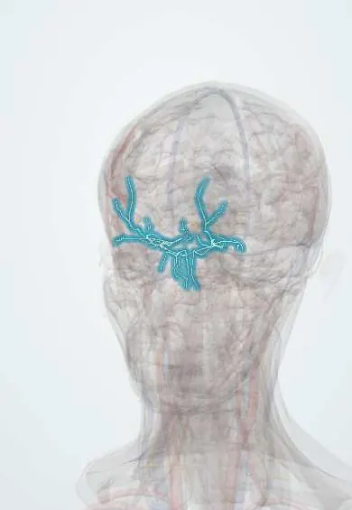
Cardiovascular system · Head and neck
Ophthalmic artery !
The ophthalmic artery, the first branch of the internal carotid inside the skull, feeds the eye, including the retina, and the orbit.
Opens in the Durgical app, no sign-up needed.
What it is
The first branch of the internal carotid, through the optic canal into the orbit.
What it does
Feeds the eye (including the central retinal artery), the orbit, the forehead and the top of the nose.
Key facts
- Origin
- Internal carotid (first branch); optic canal
- Branches
- Central retinal, lacrimal, posterior ciliary (short and long), muscular (anterior ciliary), supraorbital, anterior and posterior ethmoidal, medial palpebral; ends as the supratrochlear and dorsal nasal arteries
- Supplies
- Eye, orbit, forehead, upper nose
Relations
- Course
- In the optic canal it lies below and lateral to the optic nerve, inside its dural sheath; in the orbit it crosses over the optic nerve to the medial side and runs forward between the superior oblique and medial rectus, ending at the upper inner orbital margin
- Medial
- Along the medial wall it travels with the nasociliary nerve, giving the ethmoidal arteries into their foramina
How to find it
It is not felt. Its end branches leave the orbit at the upper margin (supraorbital notch and above the inner canthus) for the forehead, and the branches of its central retinal artery are seen directly on fundoscopy.
On imaging
Carotid Doppler, CT or MR angiography show it at its origin; orbital Doppler can show reversed flow in severe internal carotid stenosis. Its origin is the landmark for paraclinoid (ophthalmic segment) aneurysms.
Clinical notes
- Central retinal artery occlusion: sudden painless loss of vision in one eye with a pale retina and a cherry-red spot at the macula; the same embolic sources as in stroke are looked for
- Giant cell arteritis in older adults can block its posterior ciliary branches and cause permanent loss of vision (arteritic ischaemic optic neuropathy)
- An aneurysm at its origin can press on the optic nerve and cause a visual field defect
- In severe nosebleeds from high in the nose, the anterior ethmoidal artery may be tied through a small incision at the medial orbit
Anatomical variants
- It usually crosses above the optic nerve; in a minority it crosses below
- Rarely it arises from the middle meningeal artery (an external carotid branch), or the middle meningeal arises from it; this matters in embolisation procedures
Development
It comes from the internal carotid, but part of the orbit is first supplied by the stapedial artery, whose territory later passes to the middle meningeal; when that early pattern persists, the ophthalmic and middle meningeal arteries share supply or one arises from the other.
Related conditions
Test yourself
Think of the answer, then tap the question to see it.
Where does the ophthalmic artery arise, and how does it enter the orbit?
From the internal carotid as it leaves the cavernous sinus; through the optic canal, below and lateral to the optic nerve.
What are the signs of central retinal artery occlusion?
Sudden painless loss of vision in one eye with a pale retina and a cherry-red spot at the macula.
Which branches of the ophthalmic artery supply the upper nasal cavity, and why do they matter?
The anterior and posterior ethmoidal arteries: a source of nosebleeds and a landmark in endoscopic sinus surgery.
How can the ophthalmic artery act as a collateral in internal carotid occlusion?
Its links with external carotid branches (facial, superficial temporal, maxillary) let its flow reverse and feed the brain.
Educational information, not medical advice. For any symptom, see a health professional.
Its names
- Official Latin (TA2)
- arteria ophthalmica TA2 4469
- Official Spanish (DeCS)
- Arteria Oftálmica DeCS D009880
- English (TA2)
- Ophthalmic artery
Related
Sources: Terminologia Anatomica (TA2); DeCS; Anatomy, Head and Neck, Ophthalmic Arteries; Anatomy, Head and Neck, Eye Arteries; Anatomy of the Human Body (20th ed.): Development of the Vascular System (Lea & Febiger (Gray's Anatomy, 1918), 1918) · adapted, Public domain.
Spotted a mistake?We are always improving for the community. Tell us what is wrong and our team will review it.Report a mistake
Representative model, for educational purposes only.