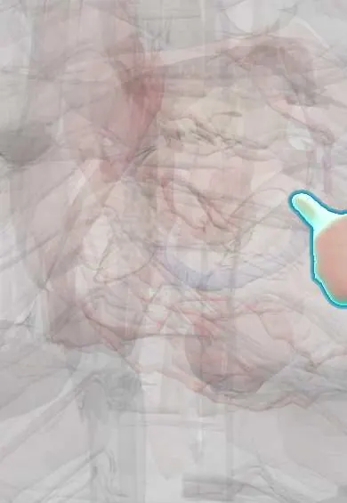
Left ventricle papillary muscles !
The papillary muscles of the left ventricle are two muscle pillars that tighten the chordae, keeping the mitral valve shut with each beat.
Official name: Superior papillary muscle of left ventricle
Opens in the Durgical app, no sign-up needed.
What it is
Two finger-like muscles (anterolateral and posteromedial) rising from the left ventricle wall, anchoring the mitral chordae.
What it does
Tighten the chordae as the ventricle squeezes, keeping the mitral valve closed.
Key facts
- Where
- Left ventricle: anterolateral and posteromedial
- Attaches
- Each sends chordae to both mitral leaflets
- Function
- Keep mitral leaflets from everting in systole; posteromedial (single supply, usually PDA) ruptures more often after infarction
Relations
- Lateral
- Anterolateral papillary muscle on the anterolateral wall, sending chordae to the lateral halves of both leaflets (anterolateral commissure)
- Below
- Posteromedial papillary muscle on the inferior wall near the septum, supplied mainly by the posterior descending artery (PDA), to the medial halves of both leaflets
- Above
- Mitral anterior leaflet and mitral posterior leaflet, through the chordae tendineae
How to find it
Opening the left ventricle shows two stout muscles rising from the middle and lower wall, one under each mitral commissure; each sends chords to both leaflets, so one muscle failing affects both.
On imaging
Echocardiography: the parasternal short axis at mid-ventricular level shows both muscles, roughly at 4 and 8 o'clock; a ruptured head appears as a mass flailing into the left atrium, clearest on transesophageal echo.
Common problems
- Rupture after a heart attack (rare, an emergency); the posteromedial one is more vulnerable because it has a single blood supply.
How it is diagnosed
Echocardiogram.
Treatment
- Surgical repair: resecting the floppy segment or adding artificial chords.
Clinical notes
- Rupture is one of the three classic mechanical complications of infarction, with free wall and septal rupture, and needs emergency surgery
- When the ventricle dilates, the muscles move apart and outward and tether the leaflets, causing functional mitral regurgitation
Anatomical variants
- The number of heads, their origin and their chordal attachments vary: each muscle may be single, bifid or multi-headed
Development
They form from the trabecular meshwork of the embryonic ventricle. If the two do not separate properly, a single muscle receives all the chords (parachute mitral valve), which narrows the valve.
Related conditions
- Mechanical complications of myocardial infarction
- Mitral regurgitation
- Myocardial Infarction (Heart Attack)
Test yourself
Think of the answer, then tap the question to see it.
Which left ventricle papillary muscle ruptures more often after infarction, and why?
The posteromedial muscle, because it has a single blood supply, usually from the posterior descending artery.
Why does the anterolateral papillary muscle of the left ventricle rarely rupture?
It has a double blood supply: diagonal branches of the LAD and marginal branches of the circumflex artery.
How does papillary muscle rupture after myocardial infarction present?
Usually in the first week: sudden acute mitral regurgitation, lung edema and shock; the murmur may be soft or absent. It needs emergency surgery.
How does a dilated left ventricle cause functional mitral regurgitation?
The papillary muscles move apart and outward and tether the mitral leaflets.
Educational information, not medical advice. For any symptom, see a health professional.
Its names
- Official Latin (TA2)
- musculus papillaris superior ventriculi sinistri; musculus papillaris inferior ventriculi sinistri TA2 4067
- Official Spanish (DeCS)
- Músculos papilares del ventrículo izquierdo DeCS D010210
- English (TA2)
- Superior papillary muscle of left ventricle
- Also called
- anterior papillary muscle of left ventricle · posterior papillary muscle of left ventricle · musculus papillaris sinister · superolateral papillary muscle
Related
- Left ventricle
- Chordae tendineae
- LAD (anterior descending artery)
- Circumflex artery
- Right coronary artery
Sources: Terminologia Anatomica (TA2); DeCS; Anatomy, Thorax, Heart Papillary Muscles; Anatomy of the Human Body (20th ed.): Development of the Vascular System (Lea & Febiger (Gray's Anatomy, 1918), 1918) · adapted, Public domain.
Spotted a mistake?We are always improving for the community. Tell us what is wrong and our team will review it.Report a mistake
Representative model, for educational purposes only.