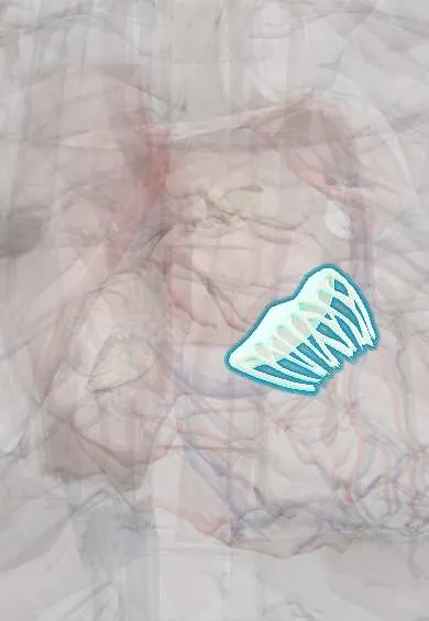
Mitral posterior leaflet !
The posterior leaflet is the smaller, back flap of the mitral valve; it closes against the anterior leaflet and is the one that most often prolapses.
Official name: Posterior leaflet of left atrioventricular valve
Opens in the Durgical app, no sign-up needed.
What it is
The longer, narrower back leaflet of the mitral valve, attached around the outer wall.
What it does
Closes against the anterior leaflet. Its middle scallop (P2) is the part that most often prolapses and is repaired.
Key facts
- Where
- Posterolateral (mural) leaflet; narrow but with long base around about 2/3 of annulus; usually 3 scallops (P1–P3)
- Attaches
- Mitral annulus; chordae tendineae from both anterolateral and posteromedial papillary muscles
- Function
- Closes against anterior leaflet in systole; P2 scallop is the commonest site of prolapse
Relations
- In front
- Mitral anterior leaflet, meeting it along the closure line
- Behind
- Posterior left ventricular wall at its hinge; just outside, the coronary sinus and the circumflex artery in the atrioventricular groove
- Above
- Posterior wall of the left atrium
- Below
- Chordae tendineae to both Left ventricle papillary muscles, plus short basal chords straight from the ventricular wall
How to find it
Its sounds are heard at the mitral area (apex, left 5th intercostal space, midclavicular line) with the person turned onto the left side. Seen from the opened left atrium, it is the long, narrow leaflet hinged on the back of the annulus, with three scallops (P1 to P3).
Examination
- Dynamic auscultation in prolapse: standing or Valsalva makes the ventricle smaller, so the click and murmur come earlier; squatting moves them later
On imaging
Transesophageal echo names its scallops P1 (lateral, near the left atrial appendage), P2 and P3 (medial); 3D echo gives the surgeon's view from the left atrium. In posterior prolapse the colour jet runs forward along the interatrial septum.
Common problems
- Mitral regurgitation (leak): from a prolapsing leaflet, a torn chord or a stretched ventricle; causes breathlessness and atrial fibrillation.
- Mitral stenosis (narrowing), mostly after rheumatic fever: still common in parts of Latin America.
- Mitral valve prolapse: a floppy leaflet that bulges back; usually mild.
How it is diagnosed
A heart murmur on exam; echocardiogram (transesophageal for detail).
Treatment
- Medicines for symptoms; regular echo follow-up for mild disease.
- Surgical repair (preferred when possible) or replacement; catheter-based clip repair for some high-risk patients.
Clinical notes
- Stitches in the posterior annulus can injure the circumflex artery and the coronary sinus, which run just outside it
- After an inferior infarct, scarred wall and the posteromedial papillary muscle can tether this leaflet, causing ischemic mitral regurgitation
Anatomical variants
- Extra scallops occur in about 30% of hearts, mostly in this leaflet (cited review)
Development
It forms from the outer rim of the atrioventricular canal, lifted off the left ventricular wall, whereas the anterior leaflet comes mainly from the fused central cushions.
Related conditions
- Mitral stenosis
- Mitral valve prolapse
- Rheumatic heart disease
- Infective endocarditis
- Mitral regurgitation
Test yourself
Think of the answer, then tap the question to see it.
Which scallop of the mitral posterior leaflet most often prolapses?
The middle scallop, P2; posterior leaflet prolapse is the type most often repaired rather than replaced.
What are the auscultatory findings of mitral regurgitation and mitral valve prolapse?
Regurgitation: a holosystolic murmur at the apex spreading to the left axilla; prolapse adds a mid-systolic click.
How do standing or Valsalva change the click and murmur of mitral valve prolapse?
They come earlier, because the ventricle gets smaller; squatting moves them later.
Which structures are at risk from stitches in the posterior mitral annulus?
The circumflex artery and the coronary sinus, which run just outside it in the atrioventricular groove.
Educational information, not medical advice. For any symptom, see a health professional.
Its names
- Official Latin (TA2)
- cuspis posterior valvae atrioventricularis sinistrae TA2 3990
- Official Spanish (DeCS)
- Cúspide posterior de la válvula mitral DeCS D008943
- English (TA2)
- Posterior leaflet of left atrioventricular valve
- Also called
- mural leaflet of left atrioventricular valve · posterior cusp of left atrioventricular valve · cuspis dorsalis valvulae bicuspidalis
Related
Sources: Terminologia Anatomica (TA2); DeCS; Anatomy, Thorax, Mitral Valve; Anatomy of the Human Body (20th ed.): Development of the Vascular System (Lea & Febiger (Gray's Anatomy, 1918), 1918) · adapted, Public domain.
Spotted a mistake?We are always improving for the community. Tell us what is wrong and our team will review it.Report a mistake
Representative model, for educational purposes only.