Muscular system · Head and neck
Genioglossus !
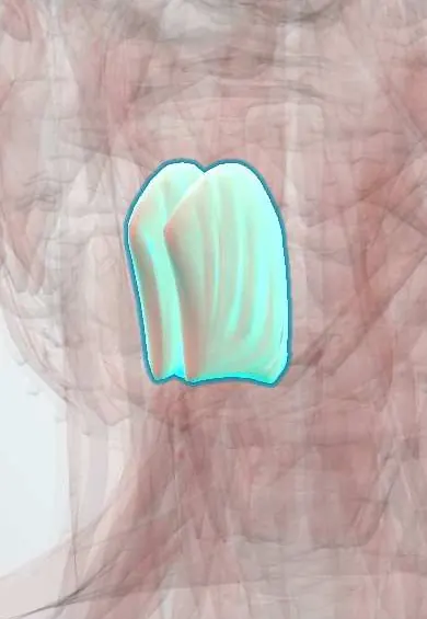
The genioglossus is the fan-shaped muscle forming most of the tongue, from the inside of the chin; it sticks the tongue out and helps keep the airway open.
Official name: Genioglossus muscle
Opens in the Durgical app, no sign-up needed.
What it is
The fan-shaped muscle that forms the bulk of the tongue, from the inside of the chin.
What it does
Sticks the tongue out; when it relaxes in sleep the tongue can fall back (sleep apnoea).
Key facts
- Origin
- Superior mental spine
- Insertion
- Body of the tongue and hyoid
- Action
- Protrudes and depresses the tongue (keeps the airway open)
- Nerve
- Hypoglossal nerve (CN XII)
- Blood supply
- Lingual artery
Relations
- Below
- Geniohyoid, then the mylohyoid
- Medial
- Its partner, separated by the fibrous lingual septum
- Lateral
- Sublingual gland and submandibular duct in front, hyoglossus behind; the lingual artery runs between the hyoglossus and the genioglossus
- Above
- Intrinsic muscles and mucosa of the tongue, into which its fibres fan out from tip to root
How to find it
Its origin can be felt as small bumps (the genial tubercles) on the inner surface of the chin; its action is seen when the person sticks the tongue straight out.
Examination
- Tongue protrusion: deviation to one side, with wasting and fasciculations on that side, points to a lower motor neuron lesion of the hypoglossal nerve on that side
- Pushing the tongue into each cheek against the examiner's finger compares the strength of the two sides
On imaging
On sagittal and coronal MRI it is the large fan of muscle from the inner chin into the tongue, on each side of the midline septum. Hypoglossal palsy shows as denervation of one half of the tongue: early oedema, later a shrunken, fatty muscle, which should prompt imaging of the whole nerve to the skull base. In sleep apnoea, sagittal MRI or a lateral cephalogram show the tongue base set back.
Clinical notes
- Hypoglossal nerve stimulation treats some cases of obstructive sleep apnoea by contracting the genioglossus during breathing in
- A fracture of the front of the mandible on both sides (flail anterior segment) can let the genioglossus pull the tongue back and block the airway
- Genioglossus advancement moves a window of inner mandible carrying the genial tubercles forward to tension the tongue base
Development
Its muscle cells come from the occipital somites and migrate forward into the tongue, drawing the hypoglossal nerve with them; this is why it is not supplied by a pharyngeal arch nerve.
Memory aid
In a lower motor neuron hypoglossal lesion, the tongue 'licks the lesion': it points to the damaged side.
Related conditions
Test yourself
Think of the answer, then tap the question to see it.
Why is the genioglossus called the airway muscle of the tongue?
Its tone holds the tongue base forward; loss of tone in sleep or unconsciousness lets the tongue fall back (obstructive sleep apnoea).
Which way does the protruded tongue deviate in a one-sided hypoglossal nerve palsy, and why?
Toward the weak side: the healthy genioglossus pushes the tongue over.
How do jaw thrust and chin lift open the airway?
They bring the mandible forward, and with it the genioglossus and the tongue.
Where does the genioglossus arise, and what supplies it?
From the superior mental spine (genial tubercles) on the inner chin; hypoglossal nerve (XII).
Educational information, not medical advice. For any symptom, see a health professional.
Its names
- Official Latin (TA2)
- musculus genioglossus TA2 2117
- Official Spanish (DeCS)
- Músculo geniogloso
- English (TA2)
- Genioglossus muscle
Related
Sources: Terminologia Anatomica (TA2); Anatomy, Head and Neck: Genioglossus Muscle; Anatomy, Head and Neck, Tongue.
Spotted a mistake?We are always improving for the community. Tell us what is wrong and our team will review it.Report a mistake
Representative model, for educational purposes only.