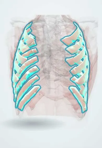
Innermost intercostals !
The innermost intercostals are the deepest layer of muscles between the ribs; the intercostal nerves and vessels run just outside them.
Official name: Innermost intercostal muscles
Opens in the Durgical app, no sign-up needed.
What it is
The deepest layer of muscle between the ribs; the intercostal nerve and vessels run just outside it.
What it does
Help steady the chest wall.
Key facts
- Origin
- Internal surface of rib above, near costal groove (lateral parts of spaces)
- Insertion
- Superior border of rib below; separated from internal intercostals by intercostal neurovascular bundle
- Action
- Probably act with internal intercostals (forced expiration); stiffen intercostal spaces
- Nerve
- Intercostal nerves (T1–T11)
- Blood supply
- Posterior and anterior intercostal arteries
Relations
- Superficial
- Intercostal nerves, posterior intercostal arteries and intercostal veins in the costal groove; then the internal intercostals
- Deep
- Endothoracic fascia, then the parietal pleura and the lung
- In front
- Transversus thoracis, on the inner side of the front chest wall
- Behind
- Subcostales near the rib angles; then the paravertebral space beside the spine
How to find it
Seen from inside the chest at thoracoscopy or dissection as a thin sheet lining the middle of each space, best developed in the lower spaces. On ultrasound it is a thin line just above the bright pleural line.
On imaging
Too thin to separate from the internal intercostals on CT or MRI. On ultrasound, the plane between the internal and innermost layers, just above the pleural line, is the target of an intercostal block, and the pleura deep to it is what the needle must avoid. Loss of lung sliding after the block suggests a pneumothorax.
Clinical notes
- Intercostal nerve block: local anaesthetic is placed under the lower border of a rib, in the plane between the internal and innermost intercostals, usually near the rib angle; pneumothorax is the main risk because the pleura lies just deeper
- Pain from the parietal pleura is felt over the chest wall in the area of the same intercostal nerves, which is why pleurisy hurts sharply with breathing
- In a paravertebral block the anaesthetic is placed beside the spine, deep to the internal intercostal membrane, where the innermost layer is absent; it reaches the nerve roots and can spread to adjacent levels
Test yourself
Think of the answer, then tap the question to see it.
Where does the intercostal neurovascular bundle lie in relation to the innermost intercostal muscles?
On their outer surface, between the innermost and the internal intercostals.
In an intercostal nerve block, where is the local anaesthetic placed and what is the main risk?
Under the lower border of a rib, between the internal and innermost intercostals, near the rib angle; pneumothorax, because the pleura lies just deeper.
Which muscles lie in the same layer as the innermost intercostals in front and behind?
The transversus thoracis in front and the subcostales behind; below, the layer continues as the plane of transversus abdominis.
Educational information, not medical advice. For any symptom, see a health professional.
Its names
- Official Latin (TA2)
- musculi intercostales intimi TA2 2313
- Official Spanish (DeCS)
- Músculos intercostales íntimos DeCS D007366
- English (TA2)
- Innermost intercostal muscles
Related
Sources: Terminologia Anatomica (TA2); DeCS; Anatomy, Thoracotomy and the Collateral Intercostal Neurovascular Bundle; Anatomy, Thorax, Wall Movements.
Spotted a mistake?We are always improving for the community. Tell us what is wrong and our team will review it.Report a mistake
Representative model, for educational purposes only.