Transversus thoracis !
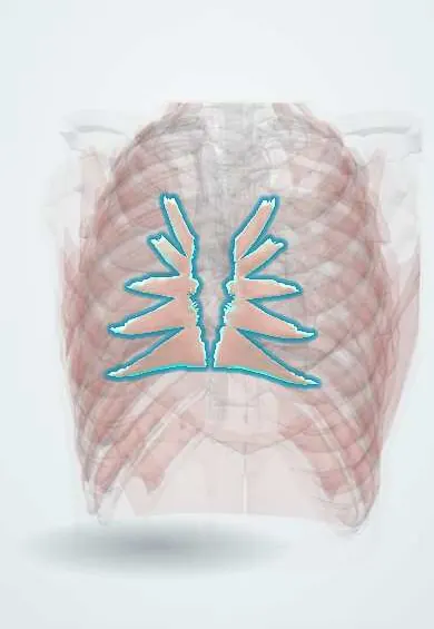
The transversus thoracis is a thin muscle on the inner surface of the front chest wall, behind the breastbone; it helps pull the ribs down.
Official name: Transversus thoracis muscle
Opens in the Durgical app, no sign-up needed.
What it is
A thin muscle on the inside of the front chest wall, behind the sternum.
What it does
Helps pull the ribs down when breathing out.
Key facts
- Origin
- Posterior surface of lower sternum (body and xiphoid process)
- Insertion
- Internal surfaces of costal cartilages 2–6
- Action
- Weakly depresses ribs and costal cartilages in expiration
- Nerve
- Intercostal nerves (T2–T6)
- Blood supply
- Internal thoracic artery (anterior intercostal branches)
Relations
- Superficial
- Internal thoracic artery and veins, parasternal lymph nodes, costal cartilages 2–6 and the internal intercostals
- Deep
- Endothoracic fascia and parietal pleura; near the midline, the pericardium in front of the heart
- Below
- Continuous with transversus abdominis at the costal margin, and close to the sternal part of the diaphragm
How to find it
Seen from inside the chest, fanning up and out from the back of the lower sternum to the costal cartilages. On parasternal ultrasound it is a thin layer under the internal thoracic vessels and above the pleural line.
On imaging
Parasternal ultrasound in a longitudinal view, a little lateral to the sternum, shows the costal cartilages, the internal thoracic vessels and, beneath them, this thin muscle over the pleura and the heart.
Clinical notes
- Internal thoracic artery harvest for coronary bypass lifts the artery off this muscle and the endothoracic fascia
- The parasternal nodes lie on it along the internal thoracic vessels; medial breast cancers can spread to them
- In a parasternal (transversus thoracis plane) block the needle stops just in front of this muscle; the internal thoracic vessels and the pleura right behind are what make ultrasound guidance standard
Anatomical variants
- Its slips vary a lot from side to side and between people: the number of digitations and the highest cartilage reached differ
Test yourself
Think of the answer, then tap the question to see it.
What are the attachments of the transversus thoracis?
From the back of the lower sternum (body and xiphoid process) to the inner surfaces of costal cartilages 2–6.
Which vessels run on the front surface of the transversus thoracis, and why does this matter in surgery?
The internal thoracic artery and veins; surgeons lift the artery off this muscle when harvesting it for coronary bypass.
Which nerves are targeted in a transversus thoracis plane block after sternotomy?
The anterior branches of the intercostal nerves, which run between the transversus thoracis and the internal intercostals.
Educational information, not medical advice. For any symptom, see a health professional.
Its names
- Official Latin (TA2)
- musculus transversus thoracis TA2 2315
- Official Spanish (DeCS)
- Músculo transverso del tórax
- English (TA2)
- Transversus thoracis muscle
Related
Sources: Terminologia Anatomica (TA2); Anatomy, Thorax, Wall Movements; Anatomy, Thoracotomy and the Collateral Intercostal Neurovascular Bundle.
Spotted a mistake?We are always improving for the community. Tell us what is wrong and our team will review it.Report a mistake
Representative model, for educational purposes only.