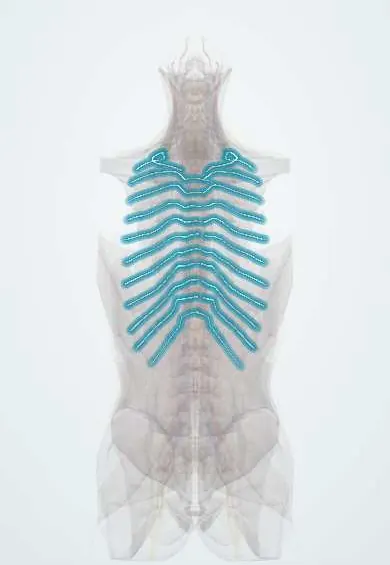
Posterior intercostal arteries !
The posterior intercostal arteries leave the thoracic aorta and run along the ribs; they feed the ribs, thoracic vertebrae and back muscles.
Opens in the Durgical app, no sign-up needed.
What it is
Small arteries from the thoracic aorta running along the ribs and into the thoracic spine.
What it does
Feed the ribs, thoracic vertebrae, back muscles and, through some branches, the spinal cord.
Key facts
- Origin
- 1st–2nd: supreme intercostal artery (costocervical trunk); 3rd–11th: thoracic aorta
- Branches
- Dorsal branch (spinal branch into the canal, back muscles, skin); collateral and lateral cutaneous branches; mammary branches from the 2nd–4th spaces to the lateral breast; anastomose with anterior intercostals
- Supplies
- Intercostal spaces, ribs, thoracic vertebrae, back muscles; thoracic cord via segmental medullary arteries
Relations
- Course
- From the back of the thoracic aorta each runs out across the vertebral body (the right ones behind the oesophagus, thoracic duct and azygos vein), behind the sympathetic trunk, into the intercostal space; near the rib angle it enters the costal groove and runs forward to meet the anterior intercostal arteries
- Above
- Posterior intercostal vein, in the costal groove of the rib above
- Below
- Intercostal nerve, lowest in the groove and least protected
- Deep
- Parietal pleura and lung, with only the innermost intercostal muscle and fascia between them
- Superficial
- External and internal intercostal muscles; near the spine, the deep back muscles
How to find it
They cannot be felt. Near the spine the artery runs across the middle of the space and is tortuous; only beyond the rib angle does it settle into the costal groove. A collateral branch runs along the upper border of the rib below.
On imaging
Not seen on plain films, but the chest radiograph shows the classic sign of aortic coarctation: notching of the lower rib borders by enlarged collaterals, sparing the first two ribs. CT angiography shows them leaving the back of the aorta and, in a haemothorax after a chest drain or rib fracture, an active contrast leak. Doppler ultrasound can find the artery before a puncture.
Clinical notes
- Injury during chest drain insertion, thoracentesis or rib fracture causes haemothorax; procedures close to the spine, where the artery lies mid-space, carry more risk
- Rib spreading in thoracotomy can injure the main and collateral neurovascular bundles, one cause of bleeding and of pain after the operation
Anatomical variants
- The supreme intercostal artery for the first two spaces usually comes from the costocervical trunk, sometimes from the thyrocervical trunk
Development
Like the lumbar arteries, they come from the segmental branches of the dorsal aorta, one pair per segment; the first two spaces are supplied from the subclavian side through the costocervical trunk instead.
Memory aid
Top to bottom in the costal groove: "VAN" (Vein, Artery, Nerve).
Related conditions
Test yourself
Think of the answer, then tap the question to see it.
What is the order of structures in the costal groove from above down, and how does it guide a chest drain?
Vein, artery, nerve (VAN); needles and chest drains pass just above the upper border of a rib to avoid the bundle.
What rib sign do enlarged posterior intercostal arteries produce in coarctation of the aorta?
Notching of the lower rib borders, sparing the first two ribs, because the arteries carry collateral flow around the narrowing.
Where do the first two posterior intercostal arteries come from, unlike the 3rd to 11th?
From the supreme intercostal artery (usually of the costocervical trunk); the 3rd to 11th come straight from the thoracic aorta.
Why do procedures close to the spine carry more risk to the posterior intercostal arteries?
Near the spine the artery runs across the middle of the intercostal space and is tortuous; it only settles into the costal groove beyond the rib angle.
Educational information, not medical advice. For any symptom, see a health professional.
Its names
- Official Latin (TA2)
- arteriae intercostales posteriores; arteria subcostalis TA2 4191
- Official Spanish (DeCS)
- Arterias intercostales posteriores
- English (TA2)
- Posterior intercostal arteries
- Also called
- arteriae intercostales
Related
- Thoracic aorta
- Artery of Adamkiewicz
- T1 vertebra
- T2 vertebra
- T3 vertebra
- T4 vertebra
- T5 vertebra
- T6 vertebra
Sources: Terminologia Anatomica (TA2); Anatomy, Thorax, Superior Intercostal Arteries; Anatomy, Thoracotomy and the Collateral Intercostal Neurovascular Bundle; Anatomy of the Human Body (20th ed.): Development of the Vascular System (Lea & Febiger (Gray's Anatomy, 1918), 1918) · adapted, Public domain.
Spotted a mistake?We are always improving for the community. Tell us what is wrong and our team will review it.Report a mistake
Representative model, for educational purposes only.