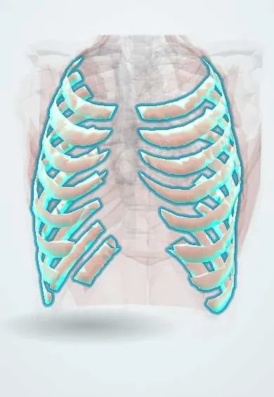
Internal intercostals !
The internal intercostals are the middle layer of muscles between the ribs; they help pull the ribs down when breathing out hard, as in coughing.
Official name: Internal intercostal muscles
Opens in the Durgical app, no sign-up needed.
What it is
Muscles between the ribs, deeper, with fibres running down and back.
What it does
Help pull the ribs down when you breathe out forcefully (coughing, blowing).
Key facts
- Origin
- Inferior border of rib above (floor of costal groove)
- Insertion
- Superior border of rib below; fibers run down and back; replaced behind by internal intercostal membrane
- Action
- Interosseous part depresses ribs in forced expiration; interchondral part elevates ribs (inspiration)
- Nerve
- Intercostal nerves (T1–T11)
- Blood supply
- Posterior and anterior intercostal arteries
Relations
- Superficial
- External intercostals; in front, between the costal cartilages, only the external intercostal membrane covers them
- Deep
- Intercostal neurovascular bundle (intercostal veins, posterior intercostal arteries and intercostal nerves), then the innermost intercostals, endothoracic fascia and pleura
- Medial
- In front they reach the sternum; behind, from the rib angles to the spine, they are replaced by the internal intercostal membrane
How to find it
Not palpable on its own. On ultrasound it is the middle of the three muscle layers between two rib shadows; the intercostal artery may be seen with colour Doppler, closer to mid-space in the back, near the spine.
On imaging
Ultrasound with a linear probe across a space shows the intercostal muscles between rib shadows, above the bright pleural line that slides with breathing; the neurovascular bundle lies under the upper rib. This view guides thoracentesis and drains over the lower rib. Contrast CT shows an intercostal artery bleeding after a drain or rib fracture.
Clinical notes
- Tube thoracostomy is usually placed in the triangle of safety (lateral border of the pectoralis major, anterior border of the latissimus dorsi and a line at nipple level, about the 5th intercostal space), going over the top of the rib
- Thoracentesis is usually done in the back or side of the chest, below the top of the effusion, again just above a rib
- A collateral branch of the bundle runs along the upper border of the rib below, so even the safe zone is not risk free; in the back the vessels lie mid-space before entering the groove
- Herpes zoster and post-thoracotomy pain follow the band of skin supplied by one intercostal nerve
Development
Derived, with the external and innermost layers, from the thoracic myotomes; their segmental origin is the reason each space has its own nerve and vessels, and why intercostal pain and weakness follow single segments.
Memory aid
VAN from top to bottom in the costal groove: Vein, Artery, Nerve.
Related conditions
Test yourself
Think of the answer, then tap the question to see it.
Between which muscle layers does the intercostal neurovascular bundle run?
Deep to the internal intercostals, between them and the innermost intercostals, sheltered in the costal groove of the rib above.
What are the borders of the triangle of safety for tube thoracostomy?
The lateral border of the pectoralis major, the anterior border of the latissimus dorsi and a line at nipple level (about the 5th intercostal space).
What is the action of the interosseous and interchondral parts of the internal intercostal muscles?
The interosseous part depresses the ribs in forced expiration; the interchondral part elevates the ribs in inspiration.
Why is even the safe zone above the rib below not risk-free when inserting a chest drain?
A collateral branch of the intercostal bundle runs along the upper border of the rib below; in the back the vessels also lie mid-space before entering the groove.
Educational information, not medical advice. For any symptom, see a health professional.
Its names
- Official Latin (TA2)
- musculi intercostales interni TA2 2312
- Official Spanish (DeCS)
- Músculos intercostales internos DeCS D007366
- English (TA2)
- Internal intercostal muscles
Related
Sources: Terminologia Anatomica (TA2); DeCS; Anatomy, Thoracotomy and the Collateral Intercostal Neurovascular Bundle; Anatomy, Thorax, Wall Movements; Anatomy of the Human Body (20th ed.): Development of the Muscles (Lea & Febiger (Gray's Anatomy, 1918), 1918) · adapted, Public domain.
Spotted a mistake?We are always improving for the community. Tell us what is wrong and our team will review it.Report a mistake
Representative model, for educational purposes only.