Cardiovascular system · Abdomen
Vessels inside the kidney !
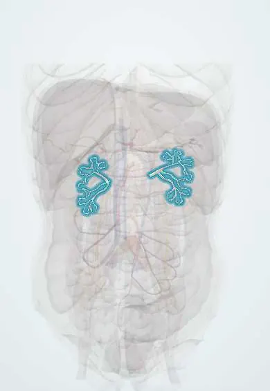
These are the arteries and veins inside the kidney; each segmental artery feeds its own zone with no backup, so a blocked branch kills a wedge of kidney.
Official name: Intrarenal arteries and veins
Opens in the Durgical app, no sign-up needed.
What it is
The branching arteries and veins inside each kidney.
What it does
Distribute blood to about a million filtering units (nephrons) per kidney.
Key facts
- Origin
- Anterior and posterior divisions of renal artery in renal sinus, giving segmental (end) arteries
- Branches
- Segmental, interlobar, arcuate, interlobular (cortical radiate), afferent arteriole, glomerulus, efferent arteriole; veins mirror back to renal vein
- Supplies
- Renal cortex and medulla (peritubular capillaries, vasa recta); segmental arteries are end arteries with no cross-flow
Relations
- Course
- Segmental arteries in the renal sinus; interlobar arteries in the renal columns between the pyramids; arcuate arteries arching over the bases of the pyramids at the corticomedullary junction; cortical radiate arteries out to the glomeruli
- Contents
- Five arterial segments: apical, anterior superior, anterior inferior and inferior from the anterior division; posterior from the posterior division
How to find it
Seen in partial nephrectomy or on a kidney cut open: the segmental arteries enter through the renal sinus, most of them in front of the renal pelvis and one posterior segmental artery behind it; their interlobar branches run in the renal columns between the pyramids and give the arcuate arteries at the border of cortex and medulla.
On imaging
Doppler of the interlobar and arcuate arteries gives a resistive index, used to follow transplants and obstruction. A slow-rising, low-peak (tardus–parvus) waveform suggests a narrowing upstream in the renal artery. On CT the corticomedullary phase shows a perfusion defect as a wedge.
Common problems
- Renal artery stenosis: a narrowing that can cause high blood pressure.
Clinical notes
- Emboli, as in atrial fibrillation or endocarditis, lodge in segmental or interlobar arteries and cause wedge-shaped infarcts
- A renal biopsy can injure an intrarenal branch, leaving an arteriovenous fistula or a pseudoaneurysm; selective embolisation of the branch controls bleeding
- The inner medulla, fed only by the vasa recta, has low oxygen levels, which helps explain renal papillary necrosis in sickle cell disease, diabetes and analgesic overuse
Anatomical variants
- Polar arteries entering the upper or lower pole directly rather than through the hilum, part of the accessory renal arteries seen in at least about 30%; each is an end artery, so tying one infarcts its territory
Related conditions
- Chronic kidney disease
- Nephrotic syndrome
- Post-streptococcal glomerulonephritis
- Diabetic kidney disease
- Lupus nephritis
- IgA nephropathy
- Hypertensive nephrosclerosis
- Polyarteritis nodosa
- Microscopic polyangiitis
- Cryoglobulinemic vasculitis
Test yourself
Think of the answer, then tap the question to see it.
What is the sequence of arteries inside the kidney, from segmental to glomerulus?
Segmental, interlobar, arcuate, interlobular (cortical radiate), afferent arteriole, glomerulus, then efferent arteriole; the veins mirror this back to the renal vein.
Why does a blocked segmental artery cause a wedge-shaped infarct?
Segmental arteries are end arteries with no cross-flow, which is also why partial nephrectomy follows segmental borders.
What is Brödel's line?
A relatively bloodless plane between the anterior and posterior arterial territories, just behind the lateral border of the kidney, used to open it.
Why can a single intrarenal vein branch be tied without harm?
Unlike the arteries, the intrarenal veins connect freely with each other.
Educational information, not medical advice. For any symptom, see a health professional.
Its names
- Official Spanish (DeCS)
- Arterias y venas intrarrenales
- English (TA2)
- Intrarenal arteries and veins
Sources: Anatomy, Abdomen and Pelvis, Renal Artery; 25.3 Gross Anatomy of the Kidney (OpenStax (Rice University), 2022) · adapted, CC BY 4.0.
Spotted a mistake?We are always improving for the community. Tell us what is wrong and our team will review it.Report a mistake
Representative model, for educational purposes only.