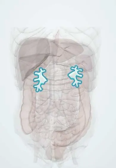
Renal pelvis !
The renal pelvis is the funnel inside each kidney that collects urine and passes it to the ureter; kidney stones often lodge where it narrows.
Opens in the Durgical app, no sign-up needed.
What it is
The funnel inside each kidney that collects urine from the calyces and narrows into the ureter.
What it does
Collects urine. Most stones form here or in the calyces.
Key facts
- Where
- Funnel in renal sinus at the hilum, most posterior of hilar structures (vein, artery, pelvis front to back)
- Function
- Collects urine and funnels it into the ureter by peristalsis
- Drains
- Minor calyces (about 7–14) into 2–3 major calyces
- Into
- Ureter at pelviureteric junction, a narrowing where stones lodge
Relations
- In front
- Renal vein and the anterior division of the renal artery with its segmental branches
- Behind
- Posterior segmental artery, which crosses behind the pelvis to the back of the kidney (the exception to VAP); then psoas major
- Below
- Narrows at the pelviureteric junction into the ureter, which runs down on psoas major
How to find it
The hilum, where the pelvis leaves the kidney, lies near the transpyloric plane from the front and at the level of the first lumbar spinous process from behind, a few centimetres from the midline. As the most posterior hilar structure it is approached from behind in pyeloplasty and percutaneous nephrolithotomy, usually through a posterior calyx.
On imaging
On ultrasound a dilated, fluid-filled pelvis with dilated calyces means hydronephrosis; an extrarenal pelvis can look similar without calyceal dilatation. CT urography opacifies the pelvicalyceal system in the excretory phase and shows filling defects (stones, clots, urothelial tumours). Non-contrast CT is the usual test for stones.
Common problems
- Kidney stones; a blockage at its outlet (UPJ obstruction).
How it is diagnosed
Blood (creatinine) and urine tests; ultrasound; CT.
Treatment
- Control blood pressure and diabetes; treat infections; dialysis or a kidney transplant for kidney failure.
Clinical notes
- A stone stuck at the pelviureteric junction causes renal colic, loin pain spreading to the groin, from stretch of the pelvis and ureter
- A staghorn calculus fills the pelvis and branches into the calyces; it is linked to infection
- Urothelial (transitional cell) carcinoma can arise in the pelvis, with the same lining as the bladder
- Pyeloplasty rebuilds an obstructed pelviureteric junction; when a vessel crosses it, the new junction is placed in front of the vessel
Anatomical variants
- Extrarenal pelvis, lying mostly outside the renal sinus
- Bifid pelvis or full duplex system, from early splitting of the ureteric bud
Development
The tip of the ureteric bud widens into the pelvis, and its branches form the major and minor calyces and the collecting ducts.
Memory aid
Renal hilum from front to back: VAP – Vein, Artery, Pelvis.
Related conditions
- Kidney stones (urolithiasis)
- Acute pyelonephritis
- Acute kidney injury
- Autosomal dominant polycystic kidney disease
- Cystinuria
- Hydronephrosis and obstructive uropathy
- Renal papillary necrosis
- Medullary sponge kidney and medullary cystic disease
Test yourself
Think of the answer, then tap the question to see it.
Where does the renal pelvis lie among the hilar structures?
It is the most posterior (vein, artery, pelvis from front to back), so it can be reached from behind without crossing the main vessels.
What are the three narrowings of the ureter where stones lodge?
The pelviureteric junction, the crossing of the iliac vessels at the pelvic brim, and the vesicoureteric junction, which is the narrowest.
What is a classic cause of pelviureteric junction obstruction in older children and adults?
A lower-pole artery crossing in front of the junction; at pyeloplasty the new junction is placed in front of the vessel.
What is a staghorn calculus?
A stone that fills the renal pelvis and branches into the calyces; it is linked to infection.
Educational information, not medical advice. For any symptom, see a health professional.
Its names
- Official Latin (TA2)
- pelvis renalis TA2 3384
- Official Spanish (DeCS)
- Pelvis renal DeCS D007682
- English (TA2)
- Renal pelvis
- Also called
- kidney pelvis · typus dendriticus · branching type
Related
Sources: Terminologia Anatomica (TA2); DeCS; Anatomy, Abdomen and Pelvis: Kidneys; Anatomy of the Human Body (20th ed.): Surface Markings of the Abdomen (Lea & Febiger (Gray's Anatomy, 1918), 1918) · adapted, Public domain; 25.3 Gross Anatomy of the Kidney (OpenStax (Rice University), 2022) · adapted, CC BY 4.0.
Spotted a mistake?We are always improving for the community. Tell us what is wrong and our team will review it.Report a mistake
Representative model, for educational purposes only.