Conditions · Nephrology and urology
Nephrotic syndrome !
Nephrotic syndrome is when damaged kidney filters leak large amounts of protein into the urine, causing swelling, low blood protein and high cholesterol.
Updated 2026-11-01
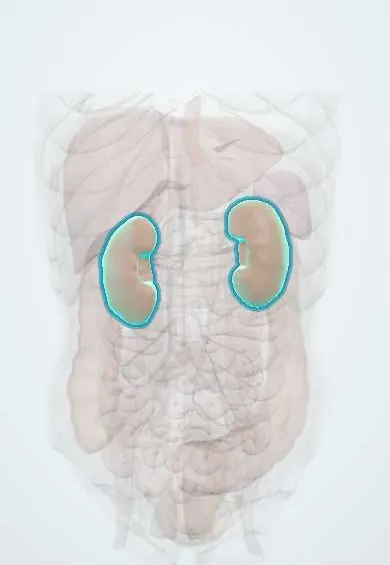
Structures involved
Tap one to see what it is and open it in 3D.
In the clinical exam
- Diagnostic facies
- Edema: pitting, grading and distribution
- Nephrotic vs nephritic syndrome
- Dehydration and fluid overload
Why it happens
- Hypercoagulable states: inherited and acquired
- Edema and effusions: the four mechanisms
- Intracellular accumulations: cholesterol, proteins, hyaline, glycogen
- Starling forces at the capillary
- Glomerular filtration: the barrier and its determinants
- Albumin
What it is
A clinical syndrome caused by damage to the glomerular filtration barrier, defined by heavy proteinuria (above about 3.5 g per day in adults), low serum albumin and oedema, usually with high blood lipids. It is a pattern with many causes, not a single disease.
Who gets it
It can appear at any age. In children it peaks between about 2 and 6 years and is slightly more common in boys, where minimal change disease is the main cause. In adults, primary glomerular diseases and secondary causes such as diabetes and lupus dominate; in Latin America, lupus nephritis and infections such as hepatitis B, hepatitis C and HIV are also considered.
Causes
- Primary (glomerular) disease: minimal change disease (most common in children), focal segmental glomerulosclerosis and membranous nephropathy (classic adult causes)
- Diabetic kidney disease, the most frequent secondary cause in adults
- Systemic diseases: lupus nephritis, amyloidosis, multiple myeloma
- Infections: hepatitis B and C, HIV, syphilis, malaria
- Drugs such as NSAIDs, and some cancers (solid tumours with membranous nephropathy, lymphoma with minimal change disease)
- Genetic podocyte disorders (congenital nephrotic syndrome)
Risk factors
- Diabetes, especially long-standing and poorly controlled
- Systemic lupus erythematosus and other autoimmune diseases
- Chronic viral infections (HBV, HCV, HIV)
- Regular use of NSAIDs
- Family history of kidney disease
Pathophysiology
Injury to the podocytes and glomerular basement membrane lets albumin pass into the urine. Low plasma albumin lowers oncotic pressure and, with renal sodium retention, fluid moves into the tissues as oedema. The liver responds by making more lipoproteins (hyperlipidaemia), and urinary loss of antithrombin and immunoglobulins causes a hypercoagulable state and a higher infection risk.
Classification
- Primary (idiopathic) versus secondary to a systemic disease, infection or drug
- By histology: minimal change disease, focal segmental glomerulosclerosis, membranous nephropathy, membranoproliferative pattern
- In children, by steroid response: steroid-sensitive, steroid-dependent, frequently relapsing or steroid-resistant
Symptoms and signs
- Swelling around the eyes (worse in the morning), legs and ankles
- Generalised oedema (anasarca), ascites or pleural effusion in severe cases
- Foamy urine
- Weight gain from retained fluid
- Fatigue and poor appetite
- Usually normal blood pressure and no visible blood in the urine in minimal change disease
Diagnosis
- Clinical
- Oedema with foamy urine; blood pressure, daily weight, signs of effusions and a search for systemic disease (rash, joint pain, diabetes history, infections, medicines).
- Tests
- Urine dipstick with heavy protein, then quantification by urine protein- or albumin-to-creatinine ratio or 24-hour urine (nephrotic range above about 3.5 g/day in adults); serum albumin low; cholesterol and triglycerides high; creatinine and eGFR. Workup for causes: glucose and HbA1c, antinuclear antibodies and complement, hepatitis B and C and HIV serology, serum and urine protein electrophoresis, and anti-PLA2R antibodies for membranous nephropathy. Kidney biopsy is the key test in adults; children with typical features are usually treated without biopsy.
- Imaging
- Kidney ultrasound (size, structure, before biopsy); Doppler ultrasound or CT when renal vein or deep vein thrombosis is suspected; chest X-ray for effusions.
Differential diagnosis
- Nephritic syndrome (haematuria, red cell casts, hypertension, less proteinuria)
- Congestive heart failure
- Liver cirrhosis with hypoalbuminaemia and ascites
- Protein-losing enteropathy or severe malnutrition
- Hypothyroidism (myxoedema)
Goal of treatment
Treat the underlying cause, reduce proteinuria and oedema, and prevent thrombosis, infection and progression to chronic kidney disease.
Treatment
- Medical
- Glucocorticoids are first-line for minimal change disease, especially in children; other immunosuppressants (calcineurin inhibitors, cyclophosphamide, rituximab, mycophenolate) are used by histological type or when steroids fail.
- Medical
- ACE inhibitors or ARBs lower proteinuria; loop diuretics with sodium restriction for oedema; statins for persistent hyperlipidaemia.
- Medical
- Anticoagulation after a thrombosis and, in selected high-risk adults (for example membranous nephropathy with very low albumin), as prevention; pneumococcal and influenza vaccination.
- Lifestyle
- Low-salt diet and adequate (not high) protein intake; fluid restriction only when needed.
- Follow-up
- Nursing care: daily weight, intake and output, oedema and skin care, urine dipstick for protein at home in children, blood pressure, and watching for infection, clots and steroid adverse effects.
Drugs for this condition
Complications
- Venous thromboembolism: renal vein thrombosis, deep vein thrombosis, pulmonary embolism
- Infections, including spontaneous bacterial peritonitis and cellulitis
- Acute kidney injury and progression to chronic kidney disease
- Hyperlipidaemia and accelerated atherosclerosis
- Hypovolaemia from aggressive diuresis, and loss of hormone-binding proteins (vitamin D, thyroid-binding globulin)
Prognosis
Depends on the cause. Most children with minimal change disease respond to steroids and keep normal kidney function, although relapses are common. Steroid-resistant forms, focal segmental glomerulosclerosis and persistent heavy proteinuria carry a higher risk of kidney failure.
Prevention
- Good control of diabetes and blood pressure
- Early treatment of lupus and of hepatitis B, hepatitis C and HIV
- Limiting nephrotoxic drugs such as NSAIDs
Test yourself
Think of the answer, then tap the question to see it.
What four findings define nephrotic syndrome?
Heavy proteinuria (above about 3.5 g/day in adults), hypoalbuminaemia, oedema and hyperlipidaemia.
What is the most common cause of nephrotic syndrome in young children?
Minimal change disease, which usually responds to glucocorticoids.
Why are people with nephrotic syndrome prone to blood clots?
Urinary loss of anticoagulant proteins such as antithrombin III, plus higher hepatic clotting factors, makes the blood hypercoagulable; renal vein thrombosis is classic.
Which test identifies the cause of nephrotic syndrome in most adults?
Kidney biopsy, supported by serology such as anti-PLA2R antibodies for membranous nephropathy.
Which drug class reduces proteinuria in most forms of nephrotic syndrome?
ACE inhibitors or angiotensin receptor blockers (ARBs).
Related conditions
- Post-streptococcal glomerulonephritis
- Diabetic kidney disease
- Systemic lupus erythematosus
- Amyloidosis
- Multiple myeloma
- HIV infection and AIDS
- Viral hepatitis
- Deep vein thrombosis
- Pulmonary embolism
- Acute kidney injury
- Chronic kidney disease
- Hyponatraemia
Educational information, not medical advice. For any symptom, see a health professional.
Sources: Nephrotic syndrome: MedlinePlus Medical Encyclopedia (MedlinePlus (U.S. National Library of Medicine), 2024); Nephrotic Syndrome in Adults (NIDDK (National Institutes of Health), 2024).
Spotted a mistake?We are always improving for the community. Tell us what is wrong and our team will review it.Report a mistake
Representative model, for educational purposes only.