Chronic kidney disease !
Chronic kidney disease is kidney damage or reduced kidney function lasting more than 3 months; it is often silent until late.
Updated 2026-10-30
Structures involved
Tap one to see what it is and open it in 3D.
- Kidneys
- Vessels inside the kidney
- Right renal artery
- Left renal artery
- Renal pelvis
- Ureters
- Abdominal aorta
In biochemistry
- Urea
- α-Galactosidase A
- LCAT (lecithin–cholesterol acyltransferase)
- Bicarbonate–CO₂ buffer system
- Cardiac troponin as a lab test
- Lipase as a lab test
- Creatinine and estimated glomerular filtration rate (eGFR)
- Urea and blood urea nitrogen (BUN) as lab tests
- Serum albumin as a lab test
- Glycated haemoglobin (HbA1c)
- Urinalysis
- Nitrogen balance
- pH, buffers and the Henderson–Hasselbalch equation
- Creatine, phosphocreatine and creatinine
- Creatinine
- Adenine phosphoribosyltransferase (APRT)
- Albumin
- Angiotensin-converting enzyme (ACE)
- Na⁺/K⁺-ATPase (sodium pump)
- Vitamin D (calcitriol)
- Iron
- Calcium and phosphate
In histology
- Kidney: cortex, medulla and the nephron
- Glomerulus and the filtration barrier: podocytes, mesangium, JGA
In embryology
- Duplicated ureter, UPJ obstruction and posterior urethral valves
- Multicystic dysplastic kidney and polycystic kidney disease
In the clinical exam
In physiology
- Anemia physiology and compensation
- Primary hemostasis and platelets
- Erythropoiesis and erythropoietin
- How edema forms
- The renin–angiotensin–aldosterone system
- The ECG in potassium and calcium disorders
- Long-term blood pressure control by the kidney
- Physiology of hypertension
- Calcium regulation: PTH, vitamin D and calcitonin
- Prolactin and its control by dopamine
- Phosphate and FGF-23
- Glomerular filtration: the barrier and its determinants
- Renal clearance
- Potassium balance
- Metabolic acidosis, the anion gap and compensation
- Functional anatomy of the nephron
- Measuring GFR and plasma flow: inulin, creatinine, PAH and filtration fraction
- Buffers and the Henderson–Hasselbalch equation
- Renal handling of H⁺ and bicarbonate
- Dehydration and fluid overload
- Physiology of acute and chronic kidney failure
- Tubular secretion of organic acids and bases
Why it happens
- Pathologic calcification: dystrophic vs metastatic
- Fibrosis in organs: TGF-β and the myofibroblast
- Heavy metals: lead, mercury, arsenic and cadmium
- How organs age: normal changes vs disease
- Health effects of climate change
What it is
Abnormal kidney structure or function lasting more than 3 months, with implications for health: an eGFR below 60 mL/min/1.73 m² or markers of kidney damage such as albuminuria.
Who gets it
Common, often silent and rising with diabetes, hypertension and ageing; these two diseases lead in Colombia and Latin America, where CKD is a public-health priority with screening of people at risk. Mesoamerican nephropathy is a regional cause of uncertain origin in hot Central American farming areas. In Lima and Tumbes, Peru (2011), 16.8% of a population-based adult sample had CKD.
Causes
- Diabetic kidney disease (the leading cause)
- Hypertensive nephrosclerosis
- Glomerulonephritis, primary or from lupus and other systemic diseases
- Autosomal dominant polycystic kidney disease and other inherited diseases
- Obstructive uropathy, recurrent stones or reflux nephropathy
- Chronic tubulointerstitial disease, including from drugs such as long-term NSAIDs
Risk factors
- Diabetes and hypertension
- Older age
- Obesity and metabolic syndrome
- Cardiovascular disease
- Family history of kidney disease
- Previous acute kidney injury
- Low birth weight and smoking
Pathophysiology
Whatever the cause, loss of nephrons makes the rest hyperfilter, which causes more scarring, so the disease tends to progress. Proteinuria itself injures the tubules. As filtration falls, the kidney fails to excrete water, salt, potassium, acid and phosphate and to make erythropoietin and active vitamin D, leading to anaemia, mineral and bone disorder and uraemia.
Classification
- KDIGO G stages by eGFR (mL/min/1.73 m²): G1 ≥90 with damage, G2 60–89, G3a 45–59, G3b 30–44, G4 15–29, G5 <15 (kidney failure)
- KDIGO A stages by urine albumin-to-creatinine ratio: A1 normal to mildly raised, A2 moderately raised, A3 severely raised
- KDIGO CGA staging: Cause, GFR and Albuminuria together set the risk (the heat map)
Symptoms and signs
- Usually no symptoms until advanced stages
- Fatigue, weakness and poor exercise tolerance (anaemia)
- Leg swelling and puffiness around the eyes
- Foamy urine (proteinuria) and nocturia
- Hypertension that is hard to control
- Itching, nausea, poor appetite and a metallic taste in advanced disease
- Confusion, cramps, restless legs and reduced urine in kidney failure
Diagnosis
- Clinical
- Often found on screening of people with diabetes or hypertension. The diagnosis needs a low eGFR or markers of damage persisting for more than 3 months; the history looks for the cause and for nephrotoxic drugs.
- Tests
- Serum creatinine with eGFR (cystatin C when creatinine is unreliable), urine albumin-to-creatinine ratio and urinalysis with sediment. Electrolytes, bicarbonate, haemoglobin, calcium, phosphate, PTH and lipids track complications. Serology or kidney biopsy when glomerular disease is suspected.
- Imaging
- Kidney ultrasound: small, echogenic kidneys suggest chronic disease; it also shows obstruction, cysts and asymmetry. Kidneys may be normal-sized or large in diabetes, polycystic disease and amyloidosis.
Differential diagnosis
- Acute kidney injury (tests are repeated over 3 months)
- Obstructive uropathy
- Renal artery stenosis
- Reduced GFR from dehydration or heart failure (prerenal)
- Low muscle mass or drugs that change creatinine without true loss of function
Goal of treatment
Slow progression, lower cardiovascular risk, manage complications and prepare in time for kidney replacement therapy or conservative care in kidney failure.
Treatment
- Medical
- Renin–angiotensin system blockade (ACE inhibitor or angiotensin receptor blocker) for albuminuria and hypertension, with blood pressure targets set by current guidelines.
- Medical
- SGLT2 inhibitors slow progression in many patients, with or without diabetes; in diabetic kidney disease a non-steroidal mineralocorticoid receptor antagonist or a GLP-1 receptor agonist may be added.
- Medical
- Treatment of complications: statins for cardiovascular risk, erythropoiesis-stimulating agents and iron for anaemia, phosphate binders and vitamin D analogues for bone and mineral disorder, alkali for acidosis, diuretics for fluid overload.
- Lifestyle
- Low-salt diet, adequate but not excessive protein, glucose and weight control, stopping smoking and avoiding nephrotoxins such as NSAIDs; drug doses are adjusted to the eGFR.
- Procedure
- Kidney replacement therapy in kidney failure: haemodialysis through an arteriovenous fistula or catheter, or peritoneal dialysis.
- Surgical
- Kidney transplantation from a living or deceased donor, the best option for suitable patients.
- Conservative
- Comprehensive conservative care without dialysis for some frail or older patients who choose it.
Drugs for this condition
- Enalapril
- Losartan
- Captopril
- Irbesartan
- Iron sucrose
- Epoetin alfa
- Darbepoetin alfa
- Empagliflozin
- Dapagliflozin
- Semaglutide
- Canagliflozin
- Furosemide
- Sodium hydrogen carbonate
- Sodium polystyrene sulfonate
- Bumetanide
- Torasemide
- Metolazone
Complications
- Cardiovascular disease, the main cause of death
- Anaemia
- CKD–mineral and bone disorder (secondary hyperparathyroidism, vascular calcification)
- Hyperkalaemia and metabolic acidosis
- Fluid overload and hypertension
- Uraemia: pericarditis, encephalopathy, bleeding tendency
- Infections and drug toxicity
Prognosis
Risk rises as eGFR falls and albuminuria rises. Many patients die of cardiovascular disease before reaching kidney failure; early treatment slows progression.
Prevention
- Screening of people with diabetes, hypertension or a family history with eGFR and urine albumin
- Good control of blood pressure and blood glucose
- Avoiding long-term NSAID use and other nephrotoxins
Test yourself
Think of the answer, then tap the question to see it.
How is chronic kidney disease defined?
Abnormal kidney structure or function for more than 3 months, such as an eGFR below 60 mL/min/1.73 m² or albuminuria.
What are the two leading causes of chronic kidney disease?
Diabetes and hypertension.
Which measurements does KDIGO use to stage chronic kidney disease?
eGFR (G1 to G5) and the urine albumin-to-creatinine ratio (A1 to A3), together with the cause.
Why do patients with chronic kidney disease become anaemic?
The failing kidney makes less erythropoietin; iron deficiency and inflammation also contribute.
Which drug classes slow progression of albuminuric chronic kidney disease?
ACE inhibitors or angiotensin receptor blockers, and SGLT2 inhibitors.
Related conditions
- Type 2 diabetes mellitus
- Arterial hypertension
- Autosomal dominant polycystic kidney disease
- Acute kidney injury
- Kidney stones (urolithiasis)
- Systemic lupus erythematosus
- Hypothyroidism
- Gynaecomastia
Educational information, not medical advice. For any symptom, see a health professional.
Sources: Chronic Kidney Disease (StatPearls (NCBI Bookshelf), 2024); Burden of chronic kidney disease in resource-limited settings from Peru: a population-based study (BMC nephrology, 2015) · adapted, CC BY 4.0.
Spotted a mistake?We are always improving for the community. Tell us what is wrong and our team will review it.Report a mistake
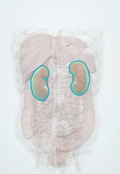
Representative model, for educational purposes only.