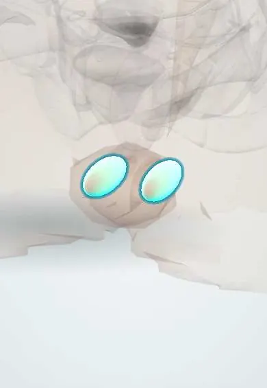
Reproductive system · Pelvis · Female
Labia majora !
The labia majora are the two outer folds of skin and fat of the vulva; they cover and protect the inner structures.
Official name: Labium majus of vulva
Opens in the Durgical app, no sign-up needed.
What it is
Two outer folds of skin and fat on either side of the vulva.
What it does
Cover and protect the inner structures; their lymph drains to the groin nodes.
Key facts
- Where
- Two folds of skin and fat from the mons pubis back toward the perineum, either side of the pudendal cleft
- Function
- Protect the vestibule; receive the end of the round ligament; homologue of the scrotum; nerves: anterior labial (ilioinguinal) and posterior labial (pudendal); lymph to superficial inguinal nodes
Relations
- Medial
- The labia minora, separated by the interlabial groove
- In front
- The mons pubis, where the two folds meet at the anterior commissure
- Behind
- The posterior commissure, in front of the perineal body and the anus
- Lateral
- The fold between the vulva and the medial thigh
- Deep
- Fat continuous with that of the mons, the end of the round ligament of the uterus, the membranous layer of superficial fascia (Colles fascia), and deeper the bulbospongiosus over the vestibular bulb
How to find it
Visible as the outer folds of the vulva from the mons pubis back to the perineum. A hernia or a canal of Nuck cyst is felt in the upper part of the labium, continuous with the superficial inguinal ring; a Bartholin cyst sits in the lower part.
On imaging
Ultrasound is the first test for a labial swelling: it shows a hernia (bowel or fat entering on straining), a canal of Nuck cyst (a fluid sac running down from the superficial inguinal ring), a Bartholin cyst lower down, or an abscess. MRI stages large vulvar cancers and their deep spread; ultrasound, CT or PET-CT look for spread to the inguinal nodes.
Common problems
- Infections and skin conditions (e.g. lichen sclerosus).
- Bartholin gland cysts and abscesses.
- Vulvar pain (vulvodynia).
- Vulvar cancer (rare, mostly in older women).
How it is diagnosed
Careful examination; swabs or a small skin biopsy when needed.
Treatment
- Depends on the cause: medicines, creams, drainage of an abscess, surgery for cancer.
Clinical notes
- After a straddle injury or a birth, blood collects easily in its loose fat; the attachments of Colles fascia stop it spreading back into the anal triangle or down the thighs
- Hair-bearing skin with sweat and sebaceous glands: a common site of folliculitis, epidermoid cysts and hidradenitis suppurativa
- Female genital mutilation/cutting type III (infibulation) narrows the vaginal opening by cutting and joining the labia minora and/or majora; it obstructs urine, menstrual flow and childbirth
Development
Forms from the labioscrotal swellings, which in the male fuse in the midline to form the scrotum.
Related conditions
- Vulvovaginal candidiasis
- Diaper dermatitis
- Chancroid
- Granuloma inguinale (donovanosis)
- Vulvar lichen sclerosus and vulvar cancer
Test yourself
Think of the answer, then tap the question to see it.
Which structure ends in the fat of the labium majus, and what labial swellings does this explain?
The round ligament of the uterus; an indirect inguinal hernia or a canal of Nuck cyst shows as a labial swelling.
To which nodes does vulvar cancer of the labia majora spread first?
The superficial inguinal nodes; lesions near the midline can drain to both sides.
Why does a pudendal block alone leave the front of the labium majus sensitive?
The front is supplied by the ilioinguinal nerve; only the back is supplied by the pudendal nerve.
Educational information, not medical advice. For any symptom, see a health professional.
Its names
- Official Latin (TA2)
- labium majus vulvae TA2 3549
- Official Spanish (DeCS)
- Labio mayor de la vulva DeCS D014844
- English (TA2)
- Labium majus of vulva
- Also called
- labium majus pudendi
Related
- Labia minora
- Superficial inguinal nodes
- Round ligament of uterus
- Pudendal nerve
- Internal pudendal artery
Sources: Terminologia Anatomica (TA2); DeCS; Anatomy, Abdomen and Pelvis: Female External Genitalia.
Spotted a mistake?We are always improving for the community. Tell us what is wrong and our team will review it.Report a mistake
Representative model, for educational purposes only.