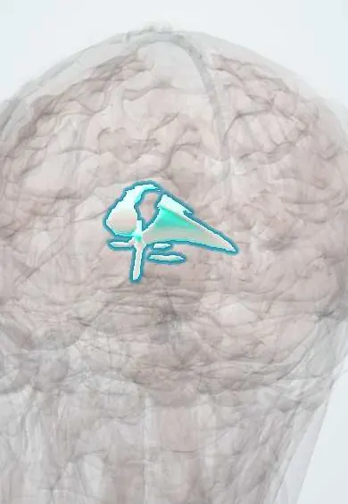
Nervous system · Head and neck
Commissures and septum !
The commissures are small fibre bridges linking the two sides of the brain; the septum is the thin wall between the front of the two lateral ventricles.
Official name: Anterior commissure
Opens in the Durgical app, no sign-up needed.
What it is
Small fibre bridges across the midline and the thin wall between the lateral ventricles.
What it does
Link the two sides; the septum separates the front of the lateral ventricles.
Key facts
- Where
- Anterior commissure in front of the fornix columns; posterior commissure above the aqueduct; septum pellucidum between the corpus callosum and fornix
- Function
- Link the two sides (temporal lobes, smell; pupillary light reflex); the septum separates the frontal horns of the lateral ventricles
Relations
- Above
- Corpus callosum, from whose underside the septum pellucidum hangs
- Below
- Body and columns of the fornix along the lower edge of the septum
- Lateral
- Frontal horns and bodies of the lateral ventricles on each side of the septum
- In front
- Lamina terminalis, the front wall of the third ventricle, which holds the anterior commissure
- Behind
- Pineal gland just above and behind the posterior commissure; the cerebral aqueduct opens beneath it
How to find it
On midsagittal MRI the anterior commissure is a small round white dot in front of the fornix columns, and the posterior commissure a smaller dot above the opening of the aqueduct, below the pineal gland. The septum pellucidum is the thin midline membrane between the frontal horns on coronal and axial images.
On imaging
A fluid space between the two leaves (cavum septi pellucidi) is normal in fetuses and newborns; its backward extension is the cavum vergae. Absence of the septum suggests septo-optic dysplasia or holoprosencephaly.
Clinical notes
- Parinaud (dorsal midbrain) syndrome: upgaze palsy, light–near dissociation of the pupils and convergence–retraction nystagmus, from a pineal tumour or hydrocephalus pressing on the region
- When only one lateral ventricle is obstructed, an endoscopic opening in the septum (septostomy) lets fluid cross to the other side
- Absence of the septum pellucidum is a feature of septo-optic dysplasia, together with small optic nerves and, in many children, pituitary hormone deficiency
Anatomical variants
- A cavum septi pellucidi persists in some adults, usually as an incidental finding; it is not part of the ventricular system (no ependymal lining, no CSF connection), so the old name "fifth ventricle" is a misnomer; a large one is more frequent after repeated head trauma, as in boxers
Development
The commissures and the septum form in the commissural plate of the lamina terminalis; the cavum between the septal leaves normally closes in the months after birth.
Test yourself
Think of the answer, then tap the question to see it.
Which fibres does the posterior commissure carry, and where does it cross?
Pretectal fibres for the consensual light reflex, crossing above the opening of the cerebral aqueduct; it is compressed in Parinaud syndrome.
What is the AC–PC line used for?
It joins the anterior and posterior commissures and is the standard reference for axial MRI slices and stereotactic surgery.
Where is the septum pellucidum, and what is measured at it on CT?
Between the corpus callosum and the fornix, forming the medial wall of each frontal horn; midline shift is measured at it.
Educational information, not medical advice. For any symptom, see a health professional.
Its names
- Official Latin (TA2)
- commissura anterior TA2 5613
- Official Spanish (DeCS)
- Comisuras y septo pelúcido DeCS D012688
- English (TA2)
- Anterior commissure
Sources: Terminologia Anatomica (TA2); DeCS; Neuroanatomy, Corpus Callosum; Tratado de anatomía humana (9.ª ed.), tomo II, Libro V, cap. VI: Cerebro (configuración exterior e interior, ventrículos, formaciones coroideas, epífisis).
Spotted a mistake?We are always improving for the community. Tell us what is wrong and our team will review it.Report a mistake
Representative model, for educational purposes only.