Joints and ligaments · Lower limb
Talocalcaneal ligaments !
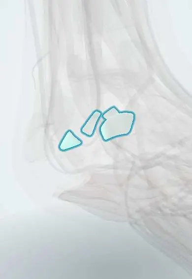
The talocalcaneal ligaments are small bands around the joint just below the ankle, holding the talus to the heel bone.
Official name: Lateral talocalcaneal ligament
Opens in the Durgical app, no sign-up needed.
What it is
Small ligaments around the subtalar joint, between the talus and the heel bone.
What it does
Hold the two bones together while the foot tilts in and out.
Key facts
- Attaches
- Lateral, medial and posterior talocalcaneal ligaments around the posterior subtalar joint
- Function
- Reinforce the subtalar joint capsule
Relations
- Lateral
- CFL superficial to the lateral talocalcaneal ligament; further out, the fibularis longus and brevis tendons
- Medial
- Flexor hallucis longus tendon passing between the talar tubercles and under the sustentaculum tali; the tibiocalcaneal fibres of the deltoid ligament lie superficial
- Behind
- Kager's fat pad and, further back, the Achilles tendon
How to find it
Not palpable as separate bands. The lateral ligament is found just in front of and deep to the calcaneofibular ligament once the fibular tendons are drawn aside; the posterior and medial ones lie either side of the flexor hallucis longus groove, as seen in posterior ankle arthroscopy.
On imaging
Too thin to assess well on routine imaging; MRI shows subtalar capsular injury and fluid. Lateral X-rays show posterior process fractures or an os trigonum at their attachments.
Clinical notes
- Fractures of the posterior process of the talus can be avulsions through these ligaments: lateral tubercle (Shepherd fracture) or medial tubercle (Cedell fracture)
- Subtalar sprains often go with lateral ankle sprains and can explain lasting hindfoot pain or giving way
- In posterior hindfoot endoscopy the flexor hallucis longus tendon is the safety line: the work stays lateral to it, because the tibial nerve and posterior tibial vessels lie just medial
Test yourself
Think of the answer, then tap the question to see it.
Which ligaments are the talocalcaneal ligaments around the posterior subtalar joint?
The lateral, medial and posterior talocalcaneal ligaments, thin bands reinforcing the capsule.
Which ligament is often torn together with the lateral talocalcaneal ligament?
The CFL, which lies just superficial to it.
What are the Shepherd and Cedell fractures?
Avulsions of the posterior process of the talus through these ligaments: lateral tubercle (Shepherd) or medial tubercle (Cedell).
Educational information, not medical advice. For any symptom, see a health professional.
Its names
- Official Latin (TA2)
- ligamentum talocalcaneum laterale; ligamentum talocalcaneum mediale; ligamentum talocalcaneum posterius TA2 1925
- Official Spanish (DeCS)
- Ligamentos talocalcáneos DeCS D013380
- English (TA2)
- Lateral talocalcaneal ligament
- Also called
- ligamentum talocalcaneare fibulare
Related
Sources: Terminologia Anatomica (TA2); DeCS; Anatomy, Bony Pelvis and Lower Limb, Foot Joints; Anatomy of the Human Body (20th ed.): Intertarsal Articulations (Lea & Febiger (Gray's Anatomy, 1918), 1918) · adapted, Public domain.
Spotted a mistake?We are always improving for the community. Tell us what is wrong and our team will review it.Report a mistake
Representative model, for educational purposes only.