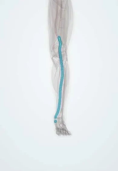
Tibial nerve !
The tibial nerve runs down the back of the knee and calf and behind the inner ankle to the sole; it powers the calf and toe flexors.
Opens in the Durgical app, no sign-up needed.
What it is
A large nerve running down the back of the knee and calf. Behind the inner ankle it passes through the tarsal tunnel and splits into the two plantar nerves.
What it does
Controls the calf muscles that point the foot and curl the toes, and, through its plantar branches, carries feeling from the sole.
Key facts
- Roots
- L4–S3
- Moves
- Posterior leg: gastrocnemius, soleus, plantaris, popliteus, tibialis posterior, FDL, FHL; sole via plantar nerves
- Feeling from
- Heel (medial calcaneal branches); sole via plantar nerves; medial sural branch to sural nerve
Relations
- Course
- Runs straight down the middle of the popliteal fossa, passing from lateral to medial behind the popliteal vessels, goes under the tendinous arch of the soleus, descends in the deep posterior compartment on the tibialis posterior with the posterior tibial artery and veins, and enters the tarsal tunnel behind the medial malleolus
- Superficial
- In the popliteal fossa, only fascia, skin and the end of the small saphenous vein; in the calf, the gastrocnemius and soleus
- Deep
- The popliteal vein and artery in the fossa; the tibialis posterior and then the tibia in the leg
- In front
- Behind the medial malleolus the posterior tibial artery and veins, with the tendons of tibialis posterior and flexor digitorum longus, lie in front of it
- Behind
- At the ankle, the flexor hallucis longus tendon lies just behind it
How to find it
In the popliteal fossa it lies in the midline just under the fascia and is shown by ultrasound superficial to the popliteal vessels. At the ankle it lies between the medial malleolus and the medial edge of the Achilles tendon, just behind the posterior tibial pulse; this is where Tinel's sign is tested and the nerve is blocked.
Examination
- Standing on tiptoe on one leg and curling the toes against resistance: inability points to the tibial nerve or the S1 root
- Tinel's sign at the tarsal tunnel: tapping behind the medial malleolus sends tingling into the sole in tarsal tunnel syndrome
- Dorsiflexion–eversion test: holding the ankle dorsiflexed and everted with the toes extended for several seconds reproduces sole symptoms in tarsal tunnel syndrome
On imaging
Ultrasound shows it in the popliteal fossa as the most superficial of the three main structures, over the popliteal vein and artery, and at the ankle just behind the posterior tibial artery. In tarsal tunnel syndrome, ultrasound or MRI look for a cause: a ganglion, varicose veins, an accessory muscle or a mass. Denervation oedema of the calf and sole muscles on MRI supports a tibial nerve lesion.
Common problems
- Injury with severe knee trauma or dislocation (uncommon).
- Compression behind the inner ankle (tarsal tunnel syndrome), causing burning or numbness in the sole.
How it is diagnosed
A physical exam; nerve conduction studies and EMG; MRI or ultrasound.
Treatment
- Depends on the cause: observation and physical therapy, or surgery to release or repair the nerve.
Clinical notes
- Knee dislocation and tibial plateau fractures can injure it together with the popliteal artery; foot pulses and sole sensation are part of every knee trauma exam
- Tarsal tunnel syndrome: compression under the flexor retinaculum by a ganglion, varicose veins, scar tissue, an accessory muscle or a valgus hindfoot
- A popliteal block catches it with the common fibular nerve; in an ankle block it is reached behind the medial malleolus, numbing most of the sole
- A tibial or sciatic nerve lesion leaves the weight-bearing sole numb, so painless pressure (trophic) ulcers of the sole are a classic late complication
Anatomical variants
- It usually divides into the plantar nerves under the flexor retinaculum, but in some feet it divides above it
- The medial calcaneal branches to the heel vary in number and origin: they may arise from the tibial nerve above the tunnel or from the lateral plantar nerve
Development
Built from the anterior divisions of the sacral plexus, it follows the ventral (flexor) muscle mass of the leg bud; the 90° medial rotation of the lower limb is why this originally ventral nerve ends up behind the knee and the leg.
Memory aid
Tarsal tunnel, front to back: 'Tom, Dick And Very Nervous Harry' (Tibialis posterior, flexor Digitorum longus, Artery, Vein, Nerve, flexor Hallucis longus).
Related conditions
- Sciatica (Lumbar Radiculopathy)
- Tarsal tunnel syndrome
- Peripheral neuropathy (diabetic and other)
- Charcot-Marie-Tooth disease
Test yourself
Think of the answer, then tap the question to see it.
What is the order of the main structures in the popliteal fossa from superficial to deep?
Tibial nerve, popliteal vein, popliteal artery; the tibial nerve is the most superficial.
Where does the tibial nerve end, and into what branches?
Under the flexor retinaculum, behind the medial malleolus, it divides into the medial and lateral plantar nerves.
What are the signs of a tibial nerve injury?
The person cannot stand on tiptoe or curl the toes, the sole is numb and the Achilles reflex (S1) is lost.
What is tarsal tunnel syndrome, and which sign supports it?
Compression of the tibial nerve under the flexor retinaculum; tapping behind the medial malleolus sends tingling into the sole (Tinel's sign).
Which tibial nerve check belongs in every knee trauma exam, and why?
Sole sensation, with foot pulses, because knee dislocation and tibial plateau fractures can injure the tibial nerve with the popliteal artery.
Educational information, not medical advice. For any symptom, see a health professional.
Its names
- Official Latin (TA2)
- nervus tibialis TA2 6582
- Official Spanish (DeCS)
- Nervio tibial DeCS D013979
- English (TA2)
- Tibial nerve
Related
- Popliteal artery
- Sciatic nerve
- Medial plantar nerve
- Lateral plantar nerve
- Flexor retinaculum
- Posterior tibial artery
- Gastrocnemius (inner head)
- Gastrocnemius (outer head)
Sources: Terminologia Anatomica (TA2); DeCS; Anatomy, Bony Pelvis and Lower Limb: Tibial Nerve; 8.5 Development of the Appendicular Skeleton (OpenStax (Rice University), 2022) · adapted, CC BY 4.0; Anatomía humana (4.ª ed.), tomo 1, cap. 70: Nervios del plexo lumbosacro.
Spotted a mistake?We are always improving for the community. Tell us what is wrong and our team will review it.Report a mistake
Representative model, for educational purposes only.