Joints and ligaments · Lower limb
CFL !
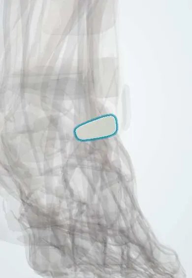
The calcaneofibular ligament runs from the outer ankle bone down to the heel; it tears in more severe ankle sprains.
Official name: Calcaneofibular ligament
Opens in the Durgical app, no sign-up needed.
What it is
A cord-like ligament on the outer ankle, from the tip of the fibula down to the heel bone.
What it does
Steadies both the ankle and the joint below it (the subtalar joint) when the foot rolls inward.
Key facts
- Attaches
- Tip of lateral malleolus to lateral surface of calcaneus, deep to fibularis tendons
- Function
- Resists inversion; stabilizes ankle and subtalar joints; second ligament torn in sprains
- Tight in
- Dorsiflexion with inversion
Relations
- Course
- From the front of the tip of the lateral malleolus downward and backward to a small tubercle on the lateral calcaneus, behind the fibular trochlea
- Superficial
- Fibularis longus tendon and fibularis brevis tendon in their common sheath, held by the fibular retinacula; the sural nerve and small saphenous vein run further back
- Deep
- Lateral talus and the posterior subtalar joint, with the thin lateral talocalcaneal ligament
- In front
- ATFL, which arises just above and in front of it
How to find it
Just below the tip of the lateral malleolus, under the fibular tendons; it is felt as a firm cord when the foot is turned in with the ankle at a right angle.
Examination
- Talar tilt: heel held, talus and calcaneus inverted with the ankle in neutral; more tilt than the other side, or a soft endpoint, suggests a CFL tear (usually with the ATFL)
- Resisted eversion with pain behind the malleolus points to the fibular tendons rather than the CFL
On imaging
Inversion stress X-rays (talar tilt) compared with the other ankle; ultrasound shows it lifting the fibular tendons when the foot dorsiflexes; MRI (coronal and axial) shows it as a dark band deep to the tendons. In the past, contrast leaking from the ankle joint into the fibular tendon sheath on arthrography meant a CFL tear.
Common problems
- Torn in more severe ankle sprains, usually after the ATFL.
- Chronic instability if it heals loose.
- The fibularis tendons run right over it and can be injured at the same time.
How it is diagnosed
A physical exam (tenderness, talar tilt test). X-rays rule out a fracture. MRI or ultrasound for ongoing problems.
Treatment
- Most sprains heal without surgery: rest, ice, compression, a brace or boot, then balance and strengthening exercises.
- Chronic instability: surgery to repair and tighten the ligament.
- If the ligament tissue is poor: reconstruction with a tendon graft from the patient or a donor.
Clinical notes
- Combined ATFL and CFL tears can leave both ankle and subtalar instability, which are hard to tell apart on examination
- Fibular tendon tears and tendon sheath problems often coexist with CFL injury and are a common cause of pain after a 'simple' sprain
- The sural nerve and fibular tendons lie close by and are at risk in lateral ligament reconstruction
Anatomical variants
- Its angle to the fibula varies widely between people, from nearly vertical to sloping well backwards
- The lower band of the ATFL is often linked to its origin by arching fibres, so the two can work as one lateral complex
Related conditions
Test yourself
Think of the answer, then tap the question to see it.
Where does the calcaneofibular ligament (CFL) attach?
From the tip of the lateral malleolus to the lateral surface of the calcaneus, deep to the fibular tendons.
Why can a calcaneofibular ligament tear loosen two joints?
It is the only lateral ankle ligament crossing two joints: the ankle and the subtalar joint.
Which test assesses the calcaneofibular ligament?
The talar tilt (inversion stress) with the ankle in neutral or slight dorsiflexion.
Which tendons lie directly over the calcaneofibular ligament and are often injured with it?
The fibularis longus and brevis tendons in their common sheath.
Educational information, not medical advice. For any symptom, see a health professional.
Its names
- Official Latin (TA2)
- ligamentum calcaneofibulare TA2 1921
- Official Spanish (DeCS)
- Ligamento calcaneofibular DeCS D017844
- English (TA2)
- Calcaneofibular ligament
- Also called
- ligamentum fibulocalcaneare
Related
Sources: Terminologia Anatomica (TA2); DeCS; Anatomy, Bony Pelvis and Lower Limb: Ankle Joint.
Spotted a mistake?We are always improving for the community. Tell us what is wrong and our team will review it.Report a mistake
Representative model, for educational purposes only.