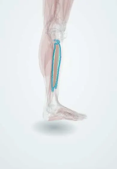
Fibularis longus !
The fibularis longus is a muscle on the outer side of the leg; it steadies the ankle, turns the foot outward and supports the arch.
Official name: Fibularis longus muscle
Opens in the Durgical app, no sign-up needed.
What it is
A muscle on the outer side of the leg.
What it does
Turns the foot outward and steadies the ankle.
Key facts
- Origin
- Head and upper two-thirds of lateral surface of fibula (some texts: also lateral condyle of tibia)
- Insertion
- Plantar base of first metatarsal and medial cuneiform (tendon crosses the sole in cuboid groove)
- Action
- Everts foot; weak plantar flexion; supports transverse arch
- Nerve
- Superficial fibular nerve (L5–S2)
- Blood supply
- Anterior tibial and fibular arteries
Relations
- Course
- From the upper lateral fibula it runs down the lateral leg, behind the lateral malleolus under the superior fibular retinaculum, below the fibular trochlea of the calcaneus, through the groove of the cuboid and across the sole
- Deep
- Fibula and fibularis brevis, which lies closer to the bone behind the malleolus; at the fibular neck, the common fibular nerve between muscle and bone
- In front
- Anterior intermuscular septum, then extensor digitorum longus in the anterior compartment
- Behind
- Posterior intermuscular septum, then soleus and flexor hallucis longus
- Below
- In the sole it runs in the deepest (fourth) layer, in a tunnel roofed by the long plantar ligament
How to find it
With the foot pushed outward against resistance, the belly firms on the upper lateral leg below the fibular head, and its tendon is felt behind the lateral malleolus, more superficial than that of fibularis brevis.
Examination
- Resisted eversion: weakness suggests a superficial or common fibular nerve lesion or an L5–S1 root problem
- Fibular tendon subluxation test: resisted eversion and dorsiflexion of the ankle; a tendon snapping forward over the malleolus suggests a torn superior fibular retinaculum
On imaging
Ankle and foot radiographs may show an os peroneum beside the cuboid; a fractured one, or one displaced up the leg, suggests a tendon rupture. Ultrasound (with dynamic views) and MRI show tears, tenosynovitis and tendon subluxation behind the lateral malleolus, where the longus lies behind and superficial to the brevis. MRI also shows split tears of the brevis next to it.
Clinical notes
- Common fibular nerve palsy at the fibular neck (fracture, tight cast, prolonged leg crossing, knee dislocation): foot drop with weak eversion and numbness over the dorsum of the foot
- Fibular tendon dislocation after a forced dorsiflexion injury, as in skiing or football, with painful snapping behind the lateral malleolus
- Painful os peroneum syndrome: lateral foot pain from a fractured sesamoid or a tear of the tendon around it
Anatomical variants
- It always reaches the base of the first metatarsal; most also attach to the medial cuneiform, and extra slips to other metatarsal bases are common
- An os peroneum is found in a minority of feet
Development
The fibular muscles come from the dorsal muscle mass, like the anterior compartment, and are supplied by the superficial branch of the same common fibular nerve.
Related conditions
Test yourself
Think of the answer, then tap the question to see it.
What is the course of the fibularis longus tendon in the foot?
Behind the lateral malleolus, round the cuboid groove and across the sole to the first metatarsal base and medial cuneiform, forming a stirrup with the tibialis anterior.
Which nerve supplies the fibularis longus, and what is its main action?
The superficial fibular nerve (L5–S2); it everts the foot and supports the transverse arch.
Which nerve winds round the fibular neck deep to the upper fibularis longus fibres, and what does its palsy cause?
The common fibular nerve; palsy causes foot drop with weak eversion and numbness of the dorsum of the foot.
What does a tendon snapping forward over the lateral malleolus on resisted eversion and dorsiflexion suggest?
Fibular tendon dislocation from a torn superior fibular retinaculum, typically after a forced dorsiflexion injury.
Educational information, not medical advice. For any symptom, see a health professional.
Its names
- Official Latin (TA2)
- musculus fibularis longus TA2 2652
- Official Spanish (DeCS)
- Músculo fibular largo
- English (TA2)
- Fibularis longus muscle
- Also called
- musculus peroneus longus · peroneus longus muscle
Related
Sources: Terminologia Anatomica (TA2); Anatomy, Bony Pelvis and Lower Limb: Calf Peroneus Longus Muscle.
Spotted a mistake?We are always improving for the community. Tell us what is wrong and our team will review it.Report a mistake
Representative model, for educational purposes only.