Joints and ligaments · Lower limb
Deltoid ligament !
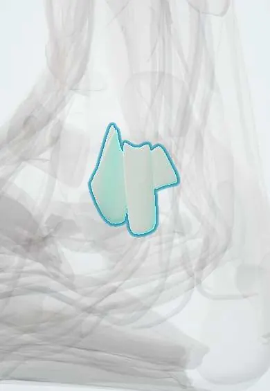
The deltoid ligament is a strong fan of ligaments on the inner ankle; it resists the foot rolling outward.
Official name: Medial collateral ligament of ankle
Opens in the Durgical app, no sign-up needed.
What it is
A strong, fan-shaped group of ligaments on the inner ankle, from the tip of the tibia to the talus, heel bone and navicular.
What it does
Holds the inner ankle together and resists the foot twisting outward. It also helps support the inner arch.
Key facts
- Attaches
- Medial malleolus fanning to navicular, spring ligament, sustentaculum tali and talus (four parts)
- Function
- Resists eversion and abduction of foot; stabilizes medial ankle; so strong it may avulse the malleolus
- Tight in
- Eversion (anterior fibers also in plantarflexion)
Relations
- Superficial
- Tibialis posterior tendon and the flexor digitorum longus tendon in their sheaths, held down by the flexor retinaculum; the tibialis posterior sheath lies directly on the ligament
- Deep
- Medial surface of the talar body and the medial gutter of the ankle joint, lined by synovium
- In front
- Great saphenous vein and saphenous nerve, crossing in front of the medial malleolus
- Below
- Spring ligament, which its tibiospring fibres join, and the sustentaculum tali of the calcaneus
How to find it
Its apex is the tip and anterior border of the medial malleolus; the fan lies in the hollow just below and in front of it. Tenderness here after a twisting injury, especially with a lateral malleolus fracture, raises suspicion of a deltoid ligament tear.
Examination
- External rotation (Kleiger) test: knee bent to 90°, ankle neutral, the foot is rotated outward; pain on the medial side suggests deltoid injury, pain over the front of the syndesmosis suggests a high ankle sprain
- Eversion (valgus) stress: medial pain and a talus that tilts or shifts more than on the other side suggest a deltoid tear
On imaging
Mortise view: the medial clear space between the medial malleolus and the talus should match the superior clear space; widening (more than about 4 mm in most texts) points to a deep deltoid tear. With an isolated fibular fracture, gravity or manual external rotation stress views or standing views test this. MRI shows both layers.
Common problems
- Sprain or tear when the foot twists outward, often together with a fibula fracture or a high ankle sprain.
- Gradual stretching in adult flatfoot.
- Tears on its own are uncommon.
How it is diagnosed
A physical exam; X-rays, sometimes taken standing or with gentle stress, to see if the ankle joint has widened. MRI.
Treatment
- Partial injuries: a boot or brace and physical therapy.
- Surgery to repair the ligament, often while fixing an ankle fracture.
- Long-standing cases: reconstruction with a tendon graft from the patient or a donor.
Clinical notes
- Ankle fracture stability depends on it: a Weber B fibular fracture with an intact deltoid ligament usually keeps the talus centred
- A torn deltoid ligament or the tibialis posterior tendon can fold into the medial gutter and block reduction of an ankle fracture-dislocation
- In late adult-acquired flatfoot the deltoid ligament gives way and the talus tilts into valgus within the mortise
- Isolated tears are rare; most deltoid injuries come with a fibular fracture or a syndesmosis injury
Anatomical variants
- Texts name its bands differently: the superficial layer is usually split into tibionavicular, tibiospring and tibiocalcaneal bands and the deep layer into anterior and posterior tibiotalar bands, and not every band is present in every ankle
Memory aid
Named after the Greek letter delta (Δ): the apex sits on the medial malleolus and the base spreads to the navicular, talus and calcaneus.
Related conditions
Test yourself
Think of the answer, then tap the question to see it.
Where does the deltoid ligament attach?
From the medial malleolus fanning to the navicular, spring ligament, sustentaculum tali and talus.
Which motion does the deltoid ligament resist?
Eversion and abduction of the foot; it is so strong it may avulse the medial malleolus.
Why does a torn deep deltoid ligament matter with a lateral malleolus fracture?
It turns a stable injury into an unstable 'bimalleolar equivalent', letting the talus shift laterally.
Which X-ray finding suggests a deep deltoid ligament tear?
A medial clear space wider than the superior clear space (more than about 4 mm in most texts) on the mortise view.
Educational information, not medical advice. For any symptom, see a health professional.
Its names
- Official Latin (TA2)
- ligamentum collaterale mediale articulationis talocruralis TA2 1913
- Official Spanish (DeCS)
- Ligamento colateral medial del tobillo (deltoideo)
- English (TA2)
- Medial collateral ligament of ankle
- Also called
- ligamentum deltoides · ligamentum mediale
Related
Sources: Terminologia Anatomica (TA2); Anatomy, Bony Pelvis and Lower Limb: Ankle Joint; Anatomy of the Human Body (20th ed.): Talocrural Articulation or Ankle-joint (Lea & Febiger (Gray's Anatomy, 1918), 1918) · adapted, Public domain.
Spotted a mistake?We are always improving for the community. Tell us what is wrong and our team will review it.Report a mistake
Representative model, for educational purposes only.