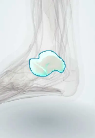
Talus !
The talus is the ankle bone, held between the tibia and fibula; it links the leg to the foot and is mostly covered in cartilage.
Opens in the Durgical app, no sign-up needed.
What it is
The ankle bone. It sits between the tibia and fibula above and the heel bone below, and joins the navicular in front.
What it does
Passes the body's weight from the leg to the foot. The foot points up and down at the joint above it, and tilts in and out at the joint below it.
Key facts
- Joints with
- Tibia and fibula (ankle); calcaneus (subtalar joint); navicular (talonavicular joint)
- Landmarks
- Head; neck; body with trochlea (dome); lateral process; posterior process with groove for flexor hallucis longus tendon; no muscle attachments
Relations
- Above
- Trochlea under the tibial plafond, with facets against the medial and lateral malleoli
- Below
- Calcaneus at the subtalar joint (posterior, middle and anterior facets); between them the sinus tarsi and tarsal canal with the interosseous talocalcaneal ligament
- In front
- Head with the navicular, cradled below by the spring ligament
- Behind
- Posterior process with medial and lateral tubercles; the flexor hallucis longus tendon runs in the groove between them
- Medial
- Deep fibres of the deltoid ligament; tibialis posterior, flexor digitorum longus, the posterior tibial artery and tibial nerve pass close behind the medial malleolus
- Lateral
- Anterior talofibular ligament from the neck and posterior talofibular ligament to the lateral tubercle; lateral process under the tip of the fibula
How to find it
With the foot plantarflexed and inverted, the front of the dome is felt just in front of the ankle joint line between the tendons. The head is felt in front of the medial malleolus, more prominent when the foot is everted; in front of the lateral malleolus the sinus tarsi forms a soft hollow.
On imaging
AP, mortise and lateral ankle views; the Canale view (foot pronated, beam tilted) shows the neck. CT for fracture planning; MRI for osteochondral lesions and avascular necrosis. On the lateral view an os trigonum is a smooth, corticated ossicle behind the posterior process.
Common problems
- Osteochondral lesions of its cartilage-covered top, often after a sprain.
- Fractures, usually from high-energy injuries. Its blood supply is fragile, so part of the bone can die (avascular necrosis).
- Fractures of its small outer or back projections, which can be mistaken for a sprain.
How it is diagnosed
X-rays; CT shows fractures in detail; MRI shows cartilage damage, bone bruising or loss of blood supply.
Treatment
- Non-displaced fractures and small lesions: a boot or cast and no weight on the foot for a while.
- Displaced fractures: surgery with screws or plates.
- Cartilage lesions: repair or grafting with bone and cartilage from the patient or a donor.
Clinical notes
- Lateral process fracture (snowboarder's fracture) mimics a lateral ankle sprain; tenderness lies just below the tip of the lateral malleolus
- Posterior ankle impingement in ballet dancers and footballers: an os trigonum or Stieda process is pinched in forced plantarflexion, often with flexor hallucis longus tendinopathy
- Subtalar dislocation and complete extrusion of the talus are high-energy injuries with a high risk of avascular necrosis
Anatomical variants
- Os trigonum, an unfused lateral tubercle of the posterior process, reported in roughly 1–25%
- Stieda process: an elongated lateral tubercle of the posterior process
- Talocalcaneal coalition (usually the middle facet), a cause of rigid flatfoot in adolescents with a C sign on the lateral X-ray
Development
Ossifies from a single centre before birth; the lateral tubercle of the posterior process has its own small centre in late childhood, which may stay separate as an os trigonum.
Related conditions
Test yourself
Think of the answer, then tap the question to see it.
Which bones does the talus articulate with?
Tibia and fibula (ankle), calcaneus (subtalar joint) and navicular (talonavicular joint).
Why are talar neck fractures at risk of avascular necrosis?
The talus is mostly cartilage with no muscle attachments, so vessels enter only through small bare areas; neck fractures cut the body's supply.
What does the Hawkins sign on X-ray 6–8 weeks after a talar neck fracture mean?
A subchondral lucency showing that the blood supply of the talar body survived.
Which injury is a lateral process fracture of the talus easily mistaken for?
A lateral ankle sprain; the snowboarder's fracture is tender just below the tip of the lateral malleolus.
Educational information, not medical advice. For any symptom, see a health professional.
Its names
- Official Latin (TA2)
- os tali TA2 1448
- Official Spanish (DeCS)
- Astrágalo DeCS D013628
- English (TA2)
- Talus
- Also called
- astragalus · astragalos · ankle bone · talar bone · talus bone
Related
- Talar cartilage
- Calcaneus
- Navicular
- Talocalcaneal interosseous ligament
- Talonavicular ligament
- Top-of-foot arteries
- ATFL
- Bifurcate ligament
Sources: Terminologia Anatomica (TA2); DeCS; Anatomy, Bony Pelvis and Lower Limb, Foot Talus.
Spotted a mistake?We are always improving for the community. Tell us what is wrong and our team will review it.Report a mistake
Representative model, for educational purposes only.