Conditions · Systemic conditions
Leishmaniasis !
Leishmaniasis is a parasitic disease spread by sandfly bites; it causes skin sores, damage to the nose and mouth lining, or illness of internal organs.
Updated 2026-10-29
Structures involved
Tap one to see what it is and open it in 3D.
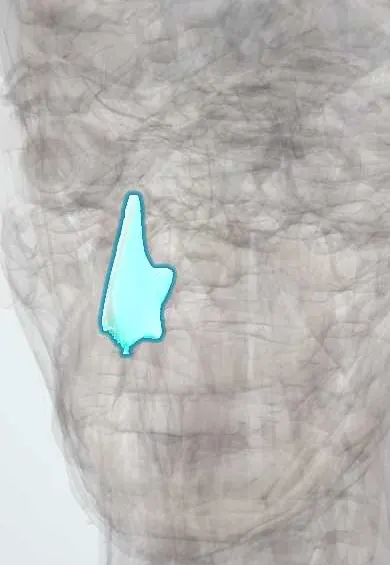
- Nasal septum
- Nasal cavity
- Spleen
- Liver
- Nasal cartilages
- Soft palate
- Pharynx
- Vocal folds
- Celiac and gastric lymph nodes
Microorganisms that cause it
In immunology
Why it happens
- Granulomatous inflammation: caseating vs non-caseating, the causes to know
- Special stains in diagnosis: Congo red, Prussian blue, PAS, Ziehl–Neelsen, Gram
- Lymph node and spleen under the microscope
What it is
A group of diseases caused by Leishmania protozoa, transmitted by the bite of infected sandflies. It affects the skin, the mucosa of the nose and mouth, or the internal organs (visceral form).
Who gets it
Present in tropical areas worldwide. In Colombia cutaneous leishmaniasis is common, linked to forest and rural work, rural tourism and military activity; the visceral form occurs in focal areas and mainly affects young children.
Causes
- Leishmania protozoa; in the Americas the Viannia subgenus (e.g. L. braziliensis, L. panamensis) causes most cutaneous and mucosal disease
- Transmission by the bite of female phlebotomine sandflies (Lutzomyia in the Americas)
- Reservoirs include wild mammals and, for visceral disease, dogs
- Rarely transfusion, needle sharing or congenital transmission
Risk factors
- Working or living in or near forests (soldiers, farmers, loggers, miners)
- Outdoor activity at dusk and night without protection
- Poor housing and nearby animal reservoirs
- Malnutrition and young age (visceral form)
- HIV and other immunosuppression
Pathophysiology
The sandfly injects promastigotes, which become amastigotes inside macrophages. In the skin, the local immune response forms a granuloma that ulcerates. Some species spread to the nasal and oral mucosa years later. In visceral disease, parasites multiply in macrophages of the spleen, liver and bone marrow, causing organ enlargement and pancytopenia.
Classification
- Cutaneous leishmaniasis: localised, disseminated or diffuse
- Mucosal (mucocutaneous) leishmaniasis, mainly in the nose, palate, pharynx and larynx
- Visceral leishmaniasis (kala-azar)
Symptoms and signs
- Cutaneous: a papule at the bite site that grows into a painless round ulcer with raised borders, on exposed skin
- Satellite lesions or nodules along the lymphatic drainage
- Mucosal: nasal stuffiness, crusting, nosebleeds, then destruction of the nasal septum and palate
- Hoarseness or difficulty swallowing if the pharynx or larynx is involved
- Visceral: prolonged fever, weight loss, a very large spleen, enlarged liver and pallor
Diagnosis
- Clinical
- A chronic painless ulcer with raised borders in someone from or returning from an endemic area. Full skin and nasal-oral examination to look for mucosal spread.
- Tests
- Cutaneous: smear (direct examination) from the ulcer border, biopsy, culture or PCR to identify the parasite and species. Visceral: bone marrow or spleen aspirate for amastigotes, rK39 serology, blood count (pancytopenia) and raised globulins. HIV testing is advised.
- Imaging
- Abdominal ultrasound to measure the spleen and liver; nasal endoscopy (by an ENT specialist) for mucosal involvement.
Differential diagnosis
- Bacterial skin ulcer or pyoderma
- Sporotrichosis and other deep fungal infections
- Cutaneous tuberculosis
- Skin cancer (basal or squamous cell carcinoma)
- Paracoccidioidomycosis or granulomatosis with polyangiitis (mucosal form)
- Malaria, lymphoma or leukaemia (visceral form)
Goal of treatment
Cure the infection, speed healing of skin lesions to limit scarring, prevent mucosal spread and, in visceral disease, prevent death.
Treatment
- Medical
- Pentavalent antimonials (e.g. meglumine antimoniate) given systemically remain a standard treatment in the Americas, with ECG, liver, pancreas and kidney monitoring.
- Medical
- Alternatives: oral miltefosine (avoided in pregnancy), pentamidine, and amphotericin B, the liposomal form being preferred for visceral and many mucosal cases.
- Procedure
- Local therapies for selected small cutaneous lesions: intralesional antimonial, thermotherapy or cryotherapy, according to national guidelines.
- Acute
- Visceral disease: treatment of anaemia, bleeding, malnutrition and secondary infections alongside antiparasitic therapy.
- Follow-up
- Follow-up after treatment to confirm healing and detect relapse or later mucosal disease.
Drugs for this condition
Complications
- Disfiguring scars
- Destruction of the nasal septum, palate and lips (mucosal disease)
- Airway obstruction or aspiration with laryngeal involvement
- Severe bacterial infections, bleeding and death in untreated visceral disease
- Drug toxicity: cardiac (QT prolongation), pancreatic, renal or hepatic
- Relapse, especially with HIV coinfection
Prognosis
Cutaneous lesions often heal with treatment, although scars remain and some species relapse or spread to the mucosa. Mucosal disease needs prolonged treatment and can leave deformity. Untreated visceral leishmaniasis is usually fatal, while treated cases mostly recover.
Prevention
- Insect repellent, long clothing and fine-mesh or insecticide-treated bed nets
- Avoiding outdoor exposure at dusk and night in endemic forest areas
- Vector control around homes
- Control of infected dogs in visceral disease foci
Test yourself
Think of the answer, then tap the question to see it.
Which insect transmits leishmaniasis?
The female phlebotomine sandfly (Lutzomyia in the Americas).
What does a typical cutaneous leishmaniasis lesion look like?
A painless round ulcer with raised, indurated borders on exposed skin, developing weeks after the bite.
Which structure is classically destroyed in mucosal leishmaniasis?
The nasal septum, with possible later spread to the palate, pharynx and larynx.
What is the clinical triad of visceral leishmaniasis?
Prolonged fever, marked splenomegaly and pancytopenia, with weight loss.
Which drug group is a standard systemic treatment for American cutaneous leishmaniasis?
Pentavalent antimonials, such as meglumine antimoniate; alternatives include miltefosine, pentamidine and amphotericin B.
Related conditions
Educational information, not medical advice. For any symptom, see a health professional.
Sources: Leishmaniasis (StatPearls (NCBI Bookshelf), 2026).
Spotted a mistake?We are always improving for the community. Tell us what is wrong and our team will review it.Report a mistake
Representative model, for educational purposes only.