Conditions · Ophthalmology and ENT
Nasal fracture and septal haematoma !
A broken nose is the most common facial fracture. A blood clot in the septum must be found and drained fast or the cartilage dies and the nose collapses.
Updated 2026-11-01
Structures involved
Tap one to see what it is and open it in 3D.
- Nasal bones
- Nasal septum
- Nasal cartilages
- Nasal cavity
- Maxillae
- Ethmoid bone
- Lacrimal bones
- Tear drainage
- Frontal sinus
Why it happens
- Hemorrhage: petechiae, purpura, ecchymosis, hematoma, and what blood loss does
- Cartilage: hyaline, elastic, fibrocartilage
What it is
A fracture of the nasal bones, often with injury to the nasal septum and cartilages, caused by a blow to the nose. Septal haematoma is a collection of blood between the septal cartilage and its lining (perichondrium); it cuts off the blood supply to the cartilage and is an emergency within the injury.
Who gets it
The nasal bones are the most frequently fractured bones of the face because they project forward and are thin. Fractures are most common in young men and result from assaults, sports, road traffic crashes (including motorcycle crashes, very frequent in Colombia) and falls. In children, the nose is more cartilaginous, so septal injuries and haematomas can occur without obvious fracture; child abuse must be considered.
Causes
- Interpersonal violence
- Contact sports (football, martial arts, boxing)
- Road traffic and motorcycle crashes
- Falls, including falls in older adults and toddlers
- Work-related injuries
Risk factors
- Young male sex
- Contact sports without face protection
- Alcohol use and exposure to violence
- Motorcycle use without a full-face helmet
- Anticoagulant use (larger haematoma) and previous nasal surgery
Pathophysiology
A lateral blow displaces the nasal bones toward the opposite side; a frontal blow flattens them and can drive force into the septum and the naso-orbito-ethmoid complex. The septal cartilage receives its nutrition from the overlying perichondrium; bleeding that lifts the perichondrium off the cartilage forms a haematoma, and without drainage the cartilage necroses within days and may become infected (septal abscess), leading to septal perforation and a saddle nose deformity.
Classification
- Undisplaced or displaced nasal bone fracture
- Simple or comminuted
- With or without septal fracture or dislocation
- Isolated or part of a more complex facial fracture (naso-orbito-ethmoid, Le Fort)
- Open (skin laceration) or closed
Symptoms and signs
- Pain, swelling and tenderness over the nasal bridge after trauma
- Nosebleed (epistaxis)
- Visible deviation or flattening of the nose
- Crepitus or mobility of the nasal bones
- Bruising around the eyes
- Nasal obstruction; with septal haematoma, a bilateral, soft, bluish-red swelling of the septum that is fluctuant to palpation
Diagnosis
- Clinical
- Clinical diagnosis: inspection for deviation, palpation for tenderness and crepitus, and mandatory anterior rhinoscopy with suction and good light to look for septal haematoma. Because swelling hides deformity, the nose is often reassessed after 3 to 7 days.
- Tests
- Testing of clear nasal fluid for beta-2 transferrin if a cerebrospinal fluid leak is suspected.
- Imaging
- Plain X-rays add little for isolated nasal fractures. CT of the face when other facial, orbital or skull base fractures are suspected.
Differential diagnosis
- Nasal contusion without fracture
- Naso-orbito-ethmoid or other midface fracture
- Orbital blowout fracture
- Basilar skull fracture with cerebrospinal fluid rhinorrhoea
- Pre-existing deviation of the nose or septum
- Septal abscess (fever and pain after an untreated haematoma)
Goal of treatment
Control bleeding, detect and drain septal haematoma, exclude associated injuries, and restore the nasal airway and shape.
Treatment
- Acute
- Assess airway and associated head, eye and facial injuries first; control epistaxis with pressure, topical vasoconstrictor and nasal packing if needed.
- Procedure
- Septal haematoma: urgent incision and drainage, then nasal packing or septal splints to prevent reaccumulation, with antibiotic cover and early follow-up.
- Conservative
- Undisplaced fractures: cold compresses, head elevation, analgesia and avoiding contact sports for several weeks.
- Procedure
- Displaced fractures: closed reduction, ideally within about 2 weeks in adults and sooner in children, before the bones set, followed by an external splint.
- Surgical
- Open reduction or later septorhinoplasty for severe septal deformity, failed closed reduction or persistent obstruction or deformity.
Complications
- Septal abscess
- Septal perforation and saddle nose deformity from cartilage necrosis
- Persistent deviation and nasal obstruction
- Impaired midfacial growth in children
- Cerebrospinal fluid leak, meningitis and cavernous sinus spread with complex injuries
- Nasolacrimal duct obstruction after naso-orbito-ethmoid fractures
Prognosis
Most isolated nasal fractures heal well; timely reduction gives good cosmetic and functional results. Missed septal haematoma is the main cause of preventable long-term deformity.
Prevention
- Face protection in contact sports
- Seat belts and full-face motorcycle helmets
- Fall prevention in older adults and young children
- Violence prevention
Test yourself
Think of the answer, then tap the question to see it.
Which finding must always be looked for inside the nose after nasal trauma?
Septal haematoma.
Why is untreated septal haematoma dangerous?
Cartilage necrosis and abscess lead to septal perforation and saddle nose deformity.
Ideal timing for closed reduction of a displaced nasal fracture in an adult?
After swelling settles, within about 2 weeks.
Clear watery rhinorrhoea after facial trauma suggests what, and which marker confirms it?
Cerebrospinal fluid leak; beta-2 transferrin.
Are plain X-rays needed for an isolated nasal fracture?
Usually not; diagnosis is clinical, CT is used for suspected complex facial fractures.
Related conditions
- Epistaxis (nosebleed)
- Orbital floor (blowout) fracture
- Basilar skull fracture
- Traumatic brain injury
- Child maltreatment (physical abuse, abusive head trauma, neglect)
- Chronic rhinosinusitis and nasal polyps
- Dacryocystitis and nasolacrimal duct obstruction
- Granulomatosis with polyangiitis
- Cocaine and sympathomimetic toxicity
Educational information, not medical advice. For any symptom, see a health professional.
Sources: Facial trauma (MedlinePlus, US National Library of Medicine, 2026).
Spotted a mistake?We are always improving for the community. Tell us what is wrong and our team will review it.Report a mistake
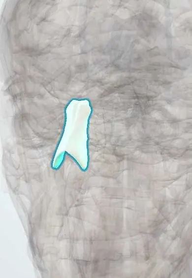
Representative model, for educational purposes only.