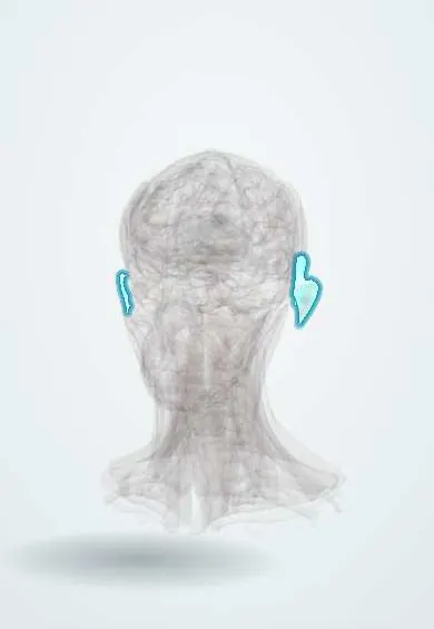
Relapsing polychondritis !
A rare autoimmune disease that inflames cartilage of the ears, nose and airway in repeated attacks and can collapse the windpipe.
Updated 2026-11-01
Structures involved
Tap one to see what it is and open it in 3D.
- Auricle
- Nasal cartilages
- Thyroid cartilage
- Nasal septum
- Cricoid cartilage
- Epiglottis
- Vocal folds
- Trachea
- Sclera
- Cochlea
- Costal cartilages
- Main bronchi
- Ascending aorta
- Valve rings (fibrous skeleton)
Why it happens
- Chronic inflammation: causes, cells and features
- Collagen
- Cartilage: hyaline, elastic, fibrocartilage
- Extracellular matrix: collagen types, elastin, ground substance
What it is
A rare immune-mediated disease with recurrent attacks of inflammation of cartilage and other tissues rich in proteoglycans: the ear, nose, larynx, trachea and bronchi, joints, eyes, inner ear and heart valves. Typical findings are a red painful ear that spares the earlobe and a saddle-nose deformity. Airway and cardiovascular involvement cause most deaths.
Who gets it
Very rare, with an estimated incidence of about 3 per million people per year. Onset is most often between 40 and 60 years, both sexes are affected about equally, and it occurs in all ethnic groups, including Latin American patients. About a third of cases occur with another disease such as a systemic vasculitis, a connective tissue disease or myelodysplastic syndrome.
Causes
- Autoimmune attack against cartilage components such as type II collagen and matrilin-1
- Genetic susceptibility (HLA-DR4)
- Association with systemic vasculitis, rheumatoid arthritis, lupus and myelodysplastic syndrome
- VEXAS syndrome: somatic UBA1 mutations in blood cells of older men that cause a relapsing polychondritis–like illness with macrocytic anaemia
Risk factors
- Age 40 to 60 years
- Other autoimmune disease
- Myelodysplastic syndrome (especially in older men)
- Possible triggers: trauma to the cartilage, such as ear piercing
Pathophysiology
Antibodies and T cells target cartilage matrix proteins. Inflammatory cells infiltrate the junction between cartilage and perichondrium, enzymes break down the matrix, and the cartilage is replaced by fibrous tissue. Loss of the cartilage framework makes the ear floppy, the nasal bridge collapse and the tracheal and bronchial walls soften (malacia) or narrow (stenosis). The same process affects the sclera, the aortic root and the cochlea.
Classification
- McAdam criteria: at least three of bilateral auricular chondritis, non-erosive seronegative polyarthritis, nasal chondritis, ocular inflammation, respiratory tract chondritis and audiovestibular damage
- Damiani modification: allows diagnosis with fewer features plus biopsy or response to treatment
- Isolated form versus forms associated with vasculitis, connective tissue disease or myelodysplasia (including VEXAS)
Symptoms and signs
- Sudden red, swollen, painful outer ear that spares the earlobe (no cartilage there)
- Nasal pain and redness, later a saddle-nose deformity
- Hoarseness, cough, wheeze or stridor from laryngotracheal disease
- Red painful eyes (episcleritis, scleritis, uveitis)
- Sudden hearing loss, tinnitus or vertigo
- Migratory non-erosive arthritis and chest wall pain at the costochondral joints
Diagnosis
- Clinical
- Diagnosis is clinical, based on typical inflammation of two or more cartilaginous sites using the McAdam or Damiani criteria. Bilateral auricular chondritis that spares the lobe is highly suggestive. Biopsy is reserved for unclear cases.
- Tests
- There is no specific blood test: ESR and CRP are raised in flares, and anaemia may be present. Blood count and smear (macrocytosis, cytopenias) and UBA1 testing in older men for VEXAS. Urinalysis and creatinine. ANCA, ANA and rheumatoid factor to look for associated diseases. Pulmonary function tests with flow-volume loops detect airway obstruction. Biopsy of affected cartilage shows chondritis.
- Imaging
- CT of the larynx and chest with inspiratory and expiratory images shows wall thickening, stenosis and malacia; bronchoscopy in selected cases. Echocardiography for the aortic valve and root. PET-CT can show active chondritis. Audiometry and eye examination in every patient.
Differential diagnosis
- Infectious perichondritis of the ear (usually unilateral, often after piercing, Pseudomonas)
- Granulomatosis with polyangiitis (saddle nose, ANCA positive, granulomas)
- Traumatic auricular haematoma (cauliflower ear)
- Congenital syphilis and leprosy (saddle nose)
- Cogan syndrome (eye and inner ear inflammation)
- Rheumatoid arthritis with scleritis
Goal of treatment
Control inflammation quickly to save the cartilage of the airway, eyes, ears and aortic root, keep long-term remission with the lowest glucocorticoid exposure, and secure the airway when it is compromised.
Treatment
- Medical
- Mild disease (ear, nose or joint only): NSAIDs, dapsone or colchicine, with short courses of glucocorticoids.
- Medical
- Moderate to severe disease (eye, airway, heart, kidney): systemic glucocorticoids plus steroid-sparing agents such as methotrexate or azathioprine, or biologics (anti-TNF agents, tocilizumab); cyclophosphamide for organ- or life-threatening disease. VEXAS needs treatment directed at the blood disorder.
- Procedure
- Procedures: airway stents, dilation of stenoses or tracheostomy for airway collapse; aortic valve or root replacement for severe regurgitation or aneurysm; hearing aids.
- Follow-up
- Monitoring: regular pulmonary function tests, airway imaging, echocardiography, audiometry, eye checks and blood counts; nursing watches for stridor and keeps airway equipment ready during flares.
Complications
- Tracheobronchial collapse or stenosis with respiratory failure and pneumonia
- Aortic regurgitation and aortic aneurysm
- Permanent sensorineural hearing loss and vestibular damage
- Saddle-nose and floppy ear deformities
- Vision loss from scleritis or uveitis
Prognosis
Course is relapsing over many years. Five-year survival has improved to about 75% to 95% with modern treatment. Outcome is worse with airway or cardiac involvement, associated systemic vasculitis and myelodysplastic syndrome or VEXAS.
Test yourself
Think of the answer, then tap the question to see it.
Which part of the ear is spared in auricular chondritis, and why?
The earlobe, because it contains no cartilage.
What are the main causes of death in relapsing polychondritis?
Airway collapse or stenosis (with infection) and cardiovascular involvement such as aortic regurgitation or aneurysm.
Which test is used to detect early airway obstruction?
Pulmonary function tests with flow-volume loops, together with inspiratory and expiratory CT.
What syndrome should be considered in an older man with polychondritis and macrocytic anaemia?
VEXAS syndrome (somatic UBA1 mutation), often associated with myelodysplasia.
Related conditions
- Uveitis
- Conjunctivitis
- Hearing loss (conductive and sensorineural, including sudden hearing loss)
- Otitis externa
- Granulomatosis with polyangiitis
- Behçet disease
- Rheumatoid arthritis
- Systemic lupus erythematosus
- Myelodysplastic syndromes
- Aortic regurgitation
- Costochondritis
- Syphilis
Educational information, not medical advice. For any symptom, see a health professional.
Sources: Relapsing Polychondritis - Symptoms, Causes, Treatment | NORD (National Organization for Rare Disorders (NORD), 2018).
Spotted a mistake?We are always improving for the community. Tell us what is wrong and our team will review it.Report a mistake
Representative model, for educational purposes only.