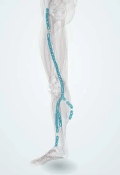
Saphenous nerve !
The saphenous nerve carries feeling from the inner knee, leg and ankle, and the inner edge of the foot; it moves no muscles.
Opens in the Durgical app, no sign-up needed.
What it is
A long skin nerve that runs beside the great saphenous vein down the inner knee and leg to the inner ankle and foot.
What it does
Carries feeling from the inner knee and leg and the inner edge of the foot.
Key facts
- Roots
- L3–L4 (longest branch of femoral nerve)
- Moves
- None (purely sensory, cutaneous)
- Feeling from
- Medial knee (infrapatellar branch), medial leg, medial ankle and medial border of foot
Relations
- Course
- Arises in the femoral triangle, enters the adductor canal lateral to the femoral artery and crosses in front of it to its medial side, leaves the canal without passing through the adductor hiatus, pierces the fascia between the sartorius and gracilis at the medial knee, then follows the great saphenous vein down the leg and in front of the medial malleolus
- Superficial
- The sartorius, which roofs the adductor canal
- Deep
- The femoral artery and vein in the canal, with the adductor longus and adductor magnus behind them; below the knee, the medial surface of the tibia
- Lateral
- The vastus medialis and the nerve to the vastus medialis, in the anterolateral wall of the adductor canal
How to find it
It becomes subcutaneous on the medial side of the knee, between the sartorius and gracilis tendons, then runs with the great saphenous vein; at the ankle it lies with the vein just in front of the medial malleolus. In the mid-thigh ultrasound finds it beside the femoral artery under the sartorius.
Examination
- Pinprick over the medial leg and medial malleolus: loss with a normal patellar reflex and normal quadriceps strength points to the saphenous nerve rather than the femoral nerve or the L4 root
- Tinel's sign where it leaves the adductor canal or at the medial knee reproduces pain or tingling in saphenous entrapment
On imaging
Too small for radiographs; ultrasound is the way to see it. In the mid-thigh it lies beside the femoral artery under the sartorius in the adductor canal, the view used for an adductor canal block. Below the knee it runs with the great saphenous vein, which is the landmark for finding and blocking it at the leg and ankle. Numbness after knee or vein surgery is a clinical diagnosis.
Clinical notes
- Injured in great saphenous vein harvest for bypass grafts, varicose vein stripping or endovenous ablation, mostly below the knee where nerve and vein run closest
- The anteromedial ankle arthroscopy portal and medial malleolus fixation can cut it
- The adductor canal block is often chosen after knee replacement because it keeps more quadriceps strength than a femoral nerve block
- Entrapment where it leaves the adductor canal can cause medial knee pain that mimics a meniscal problem
Anatomical variants
- Where it leaves the adductor canal varies: usually between the sartorius and gracilis tendons at the knee, but it may pass through the sartorius or come out in front of it
- Below the knee it may lie in front of or behind the great saphenous vein, and often splits into two or more branches that wind round the vein
Development
Its skin area on the medial leg is the L4 dermatome. Because the lower limb turns medially during week 7, the dermatomes spiral down the leg, and L4 comes to lie on the medial side down to the medial malleolus.
Related conditions
Test yourself
Think of the answer, then tap the question to see it.
Which is the only branch of the femoral nerve that reaches below the knee?
The saphenous nerve, which is purely sensory.
Why is the adductor canal block chosen after knee replacement?
It targets the saphenous nerve in the adductor canal and keeps more quadriceps strength than a femoral nerve block.
Why can great saphenous vein harvest or stripping leave the medial leg numb?
Below the knee the saphenous nerve runs with the great saphenous vein, so vein surgery can injure it.
Educational information, not medical advice. For any symptom, see a health professional.
Its names
- Official Latin (TA2)
- nervus saphenus TA2 6525
- Official Spanish (DeCS)
- Nervio safeno
- English (TA2)
- Saphenous nerve
Related
Sources: Terminologia Anatomica (TA2); Anatomy, Bony Pelvis and Lower Limb: Saphenous Nerve, Artery, and Vein; 8.5 Development of the Appendicular Skeleton (OpenStax (Rice University), 2022) · adapted, CC BY 4.0.
Spotted a mistake?We are always improving for the community. Tell us what is wrong and our team will review it.Report a mistake
Representative model, for educational purposes only.