Joints and ligaments · Lower limb
MPFL !
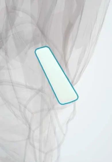
The medial patellofemoral ligament (MPFL) is the main restraint that keeps the kneecap from sliding outward; it tears in dislocations.
Official name: Medial patellofemoral ligament
Opens in the Durgical app, no sign-up needed.
What it is
A thin, flat ligament from the inner border of the kneecap to the inner side of the femur, near the adductor tubercle.
What it does
Is the main restraint that stops the kneecap from sliding out of its groove toward the outside of the knee.
Key facts
- Attaches
- Medial femur between adductor tubercle and medial epicondyle to upper medial border of patella
- Function
- Main restraint to lateral patellar displacement (about 50–60%); usually torn in patellar dislocation
- Tight in
- Knee extension and early flexion (0–30°)
Relations
- Course
- A fan-shaped band in layer 2 of the medial knee, running forward from the groove between the adductor tubercle and the medial epicondyle to the upper medial border of the patella
- Superficial
- Vastus medialis obliquus, which blends with it near the patella, and the superficial fascia (layer 1)
- Deep
- Joint capsule (layer 3) and synovium over the medial trochlea
- Above
- Adductor magnus tendon ending on the adductor tubercle, just above and behind its femoral attachment
How to find it
It cannot be felt as a separate cord. Its femoral attachment lies in the dip between the adductor tubercle (found by following the adductor magnus tendon down to its bump) and the medial epicondyle; tenderness here after a dislocation is the Bassett sign.
Examination
- Patellar apprehension test (Fairbank): knee at 20–30° of flexion, patella pushed laterally; fear or a reflex quadriceps contraction means lateral instability
- Lateral patellar glide: more than about half the patellar width, or a soft end point, compared with the other knee suggests MPFL insufficiency
- J sign: the patella jumps laterally as the knee reaches full extension, a sign of maltracking, often with trochlear dysplasia or patella alta
On imaging
MRI after a dislocation: MPFL disruption or oedema at the femoral or patellar end, bone bruises on the medial patellar facet and the lateral femoral condyle, osteochondral fragments. Risk factors are measured: trochlear dysplasia (crossing sign on the lateral X-ray), patella alta (Insall–Salvati ratio above 1.2) and TT–TG distance on CT or MRI.
Common problems
- Torn in most first-time kneecap dislocations (the kneecap slides out to the side).
- After a tear the kneecap may keep slipping out (patellar instability).
How it is diagnosed
Physical exam (the kneecap moves too far outward; the patient fears it will slip); X-rays and MRI show the tear and any bone or cartilage damage.
Treatment
- First dislocation: often a brace and physical therapy to strengthen the thigh muscles.
- Repeated dislocations: MPFL reconstruction, replacing the ligament with a tendon graft from the patient or a donor.
Clinical notes
- Reconstruction usually uses a semitendinosus or gracilis graft (or a quadriceps tendon strip), fixed to the patella and to a femoral tunnel located on a true lateral X-ray (Schöttle point)
- A femoral tunnel placed too high or too far forward makes the graft tight in flexion and overloads the medial patellofemoral joint
- In children the femoral attachment lies just below the distal femoral growth plate, which must be avoided when drilling
- Marked bony risk factors may need an added tibial tubercle osteotomy or trochleoplasty
Anatomical variants
- Width and exact attachments vary a great deal; texts disagree on whether the femoral attachment centres on the adductor tubercle, the medial epicondyle or the groove between them
Related conditions
Test yourself
Think of the answer, then tap the question to see it.
What are the attachments of the medial patellofemoral ligament (MPFL)?
From the medial femur between the adductor tubercle and the medial epicondyle, to the upper medial border of the patella.
In which range of knee flexion does the MPFL matter most, and why?
The first 20–30°, before the patella engages the trochlear groove; deeper, the bony groove holds the patella.
How is the patellar apprehension test (Fairbank) performed?
With the knee at 20–30° of flexion the patella is pushed laterally; fear or a reflex quadriceps contraction means lateral instability.
Which factors make recurrent patellar dislocation more likely after an MPFL tear?
Trochlear dysplasia, patella alta, a lateralized tibial tubercle and a first dislocation at a young age.
What is the standard operation for recurrent lateral patellar instability?
MPFL reconstruction, usually with a semitendinosus or gracilis graft fixed to the patella and a femoral tunnel.
Educational information, not medical advice. For any symptom, see a health professional.
Its names
- Official Latin (TA2)
- ligamentum patellofemorale mediale TA2 1903
- Official Spanish (DeCS)
- Ligamento patelofemoral medial
- English (TA2)
- Medial patellofemoral ligament
Related
Sources: Terminologia Anatomica (TA2); Anatomy, Bony Pelvis and Lower Limb: Medial Patellofemoral Ligament.
Spotted a mistake?We are always improving for the community. Tell us what is wrong and our team will review it.Report a mistake
Representative model, for educational purposes only.