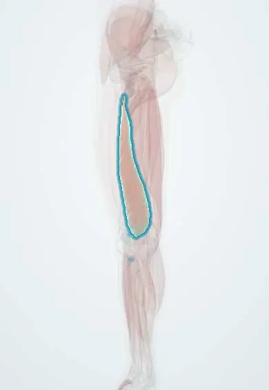
Vastus medialis !
The vastus medialis is the inner quadriceps muscle, the bulge above the inner knee; it straightens the knee and steadies the kneecap.
Official name: Vastus medialis muscle
Opens in the Durgical app, no sign-up needed.
What it is
The inner quadriceps muscle, forming the teardrop bulge above the inner knee.
What it does
Straightens the knee and helps keep the kneecap centred.
Key facts
- Origin
- Lower intertrochanteric line; spiral line; medial lip of linea aspera; medial supracondylar line; adductor magnus tendon
- Insertion
- Medial patella via quadriceps tendon; tibial tuberosity via patellar ligament; medial patellar retinaculum
- Action
- Extends knee; lower oblique fibres (VMO) resist lateral drift of patella
- Nerve
- Femoral nerve (L2–L4)
- Blood supply
- Femoral and deep femoral arteries; descending genicular artery
Relations
- Superficial
- Sartorius over the adductor canal in the middle third of the thigh; lower down only fascia and skin over the teardrop bulge
- Medial
- Adductor canal, which carries the femoral vessels, the saphenous nerve and the nerve to vastus medialis
- Behind
- Adductor longus and adductor magnus; the vastoadductor membrane stretches from it to them and closes the canal
- Lateral
- Rectus femoris in front and vastus intermedius deep, with which it partly blends
- Deep
- Near the knee, the medial patellofemoral ligament runs deep to its lowest fibres
How to find it
With the knee fully straight and the thigh tightened, or in the last degrees of a straight-leg raise, the teardrop bulge appears above and medial to the patella; its oblique lower fibres can be traced to the medial edge of the patella.
Examination
- Patellar apprehension test: with the knee slightly bent, the examiner pushes the patella laterally; guarding or fear of dislocation points to lateral patellar instability, where the restraint from these fibres and the medial patellofemoral ligament falls short
On imaging
Axial MRI and ultrasound of the lower thigh show it on the medial side. In the mid-thigh, ultrasound shows it forming the lateral wall of the adductor canal next to the femoral artery under the sartorius: the view used for an adductor canal block. After knee injury or surgery, wasting shows as a thinner muscle on ultrasound and, if long-standing, fatty change on MRI.
Common problems
- Strains (pulled muscle) from sprinting or kicking; bruising from a direct blow.
- Weakness and shrinking after knee injury or surgery, which rehabilitation works to rebuild.
How it is diagnosed
Physical exam; ultrasound or MRI for significant injuries.
Treatment
- Rest, ice, then progressive strengthening with a physical therapist.
Clinical notes
- Thigh girth measured at a fixed distance above the patella is a simple way to follow its wasting and recovery
- Strengthening its oblique fibres is a traditional focus of rehabilitation for patellofemoral pain and instability, although truly selective activation is debated
- In lateral patellar dislocation the medial patellofemoral ligament, deep to its lower fibres, is the main passive restraint torn
Development
Part of the quadriceps, formed from the dorsal muscle mass of the limb bud and supplied by the femoral nerve, as are the other vasti.
Related conditions
Test yourself
Think of the answer, then tap the question to see it.
Which quadriceps muscle wastes early and visibly after a knee injury, effusion or surgery, and how does it show?
The vastus medialis: its teardrop bulge above and medial to the patella flattens; thigh girth above the patella is used to follow it.
What role do the lower oblique fibres of the vastus medialis (VMO) play at the patella?
They resist lateral drift of the patella, balancing the lateral pull of the vastus lateralis.
Which canal does the vastus medialis help to form, and which wall?
The adductor canal: the vastus medialis forms its anterolateral wall, next to the femoral artery and vein and the saphenous nerve.
Which ligament, deep to the lowest vastus medialis fibres, is the main passive restraint torn in lateral patellar dislocation?
The medial patellofemoral ligament.
Educational information, not medical advice. For any symptom, see a health professional.
Its names
- Official Latin (TA2)
- musculus vastus medialis TA2 2620
- Official Spanish (DeCS)
- Músculo vasto medial DeCS D052097
- English (TA2)
- Vastus medialis muscle
- Also called
- musculus vastus tibialis
Related
Sources: Terminologia Anatomica (TA2); DeCS; Anatomy, Bony Pelvis and Lower Limb: Thigh Quadriceps Muscle.
Spotted a mistake?We are always improving for the community. Tell us what is wrong and our team will review it.Report a mistake
Representative model, for educational purposes only.