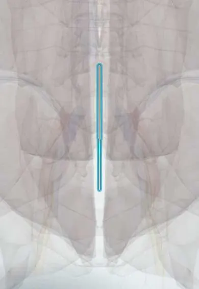
Median sacral artery !
The median sacral artery is a small artery running down the middle of the front of L5 and the sacrum; it feeds the sacrum and coccyx.
Opens in the Durgical app, no sign-up needed.
What it is
A small artery running down the middle of the front of L5 and the sacrum.
What it does
Feeds the sacrum and coccyx. It is tied off to reach the L5-S1 disc from the front.
Key facts
- Origin
- Posterior aspect of the abdominal aorta just above its bifurcation
- Branches
- Small fifth lumbar arteries; lateral sacral twigs; ends at the coccyx (coccygeal body)
- Supplies
- L5 vertebra, sacrum, coccyx, posterior rectum; ligated in anterior L5–S1 approach
Relations
- Course
- From the back of the aorta just above the bifurcation it descends in the midline over the L4 and L5 bodies, the sacrum and the coccyx, ending at the coccygeal body
- In front
- Left common iliac vein, the superior hypogastric plexus and, lower down, the rectum and its mesentery
- Behind
- L4 and L5 bodies, the L5–S1 disc and the front of the sacrum and coccyx
- Lateral
- Common iliac vessels above; lower, the sympathetic trunks and the lateral sacral vessels on the front of the sacrum
How to find it
It cannot be felt. In the anterior approach to L5–S1 it is seen in the midline on the front of the disc, between the common iliac veins, usually with a median sacral vein that drains into the left common iliac vein.
On imaging
A small vessel, usually seen only on contrast CT or CT angiography as a thin midline line from the back of the aortic bifurcation running down in front of L5 and the sacrum. Before an anterior approach to L5–S1, CT or MRI shows it and its vein in front of the disc. In a sacrococcygeal teratoma it can be enlarged as a main feeding vessel, seen on Doppler ultrasound.
Clinical notes
- Presacral dissection in rectal surgery can injure it or the nearby presacral veins
- It can enlarge as a collateral when the aorta or the iliac arteries are blocked
- In removing a large sacrococcygeal teratoma, the artery is controlled early, by tying it or by embolization before surgery, to limit bleeding
Development
It is the remnant of the caudal continuation of the dorsal aorta, which shrinks as the embryonic tail regresses.
Test yourself
Think of the answer, then tap the question to see it.
Where does the median sacral artery arise?
From the back of the abdominal aorta just above its bifurcation; it is the true end of the aorta.
Why is the median sacral artery clipped in the anterior approach to L5–S1?
It lies with its vein on the front of the L5–S1 disc, in the midline window, among the fibres of the superior hypogastric plexus.
Which newborn tumour does the median sacral artery typically feed?
Sacrococcygeal teratoma, the most common tumour of the newborn.
Educational information, not medical advice. For any symptom, see a health professional.
Its names
- Official Latin (TA2)
- arteria sacralis mediana; vena sacralis mediana TA2 4298
- Official Spanish (DeCS)
- Arteria sacra media
- English (TA2)
- Median sacral artery
- Also called
- medial sacral vein · arteria sacralis media · aorta caudalis
Related
Sources: Terminologia Anatomica (TA2); Anatomy, Abdomen and Pelvis: Abdominal Aorta.
Spotted a mistake?We are always improving for the community. Tell us what is wrong and our team will review it.Report a mistake
Representative model, for educational purposes only.