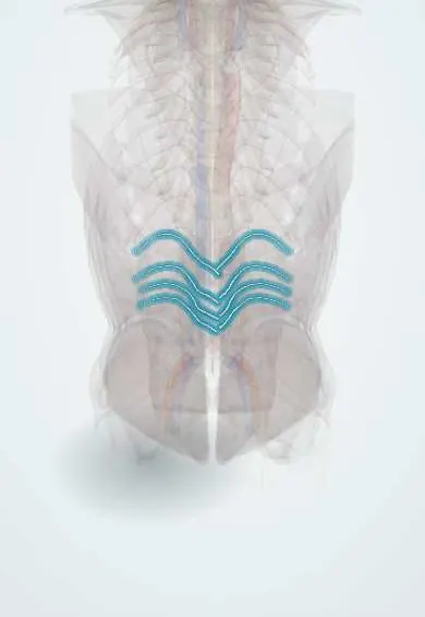
Lumbar arteries !
The lumbar arteries are pairs of small arteries from the back of the aorta that wrap each lumbar vertebra; they feed the spine and back muscles.
Opens in the Durgical app, no sign-up needed.
What it is
Pairs of small arteries from the back of the aorta that wrap around each lumbar vertebral body.
What it does
Feed the vertebrae, back muscles, nerve roots and part of the spinal cord's blood supply.
Key facts
- Origin
- Posterior aspect of the abdominal aorta; 4 pairs opposite L1–L4 (fifth pair, if present, from median sacral)
- Branches
- Dorsal branch (spinal branch into the canal, back muscles, skin); abdominal wall branches that anastomose with the lower intercostal, subcostal, inferior epigastric, iliolumbar and deep circumflex iliac arteries; radicular and segmental medullary arteries
- Supplies
- Lumbar vertebrae, cauda equina and conus, posterior abdominal wall, back muscles
Relations
- Course
- From the back of the aorta each runs around the side of a lumbar vertebral body, passes under the psoas major and mostly behind the quadratus lumborum, and at the intervertebral foramen gives its dorsal branch before continuing in the abdominal wall
- In front
- Sympathetic trunk; on the right, the inferior vena cava; the upper ones also pass behind the crura of the diaphragm
- Behind
- Vertebral bodies, at their concave mid-portion
- Lateral
- Psoas major, under whose fibrous arches they pass, then the quadratus lumborum
How to find it
They cannot be felt. In lateral (transpsoas) or anterior approaches they are seen crossing the middle of each vertebral body with their veins; on CT angiography they are paired small vessels leaving the back of the aorta at each level.
On imaging
CT angiography shows them leaving the back of the aorta in pairs and curving around the waist of each lumbar vertebral body. After endovascular aneurysm repair, contrast entering the sac from a lumbar artery (or the inferior mesenteric) is a type II endoleak, seen on CT angiography or contrast ultrasound. In trauma or bleeding on anticoagulants, CT may show a blush in a retroperitoneal haematoma.
Clinical notes
- Injury in lateral lumbar fusion, or from a pedicle screw that breaches the side of the body, can cause a retroperitoneal haematoma or a pseudoaneurysm
- An upper lumbar artery can give the artery of Adamkiewicz, so segmental vessels at L1–L2 are handled with particular care
- Their spinal branches supply the cauda equina and nerve roots and connect with one another inside the canal
Anatomical variants
- Usually four pairs come from the aorta; a small fifth pair occasionally arises from the median sacral artery, and the iliolumbar artery takes over the L5 level
Development
They are the persisting segmental branches of the embryonic dorsal aorta, which explains their regular one-pair-per-level pattern.
Related conditions
Test yourself
Think of the answer, then tap the question to see it.
How many pairs of lumbar arteries are there, and where do they arise?
Four pairs from the back of the abdominal aorta, opposite L1–L4; a fifth pair, if present, comes from the median sacral artery.
Where are the lumbar arteries tied in anterior and lateral lumbar surgery, and why?
At the middle of the vertebral body, away from the foramen, to keep the collaterals to the spinal cord and roots.
Which type of endoleak after endovascular aortic aneurysm repair comes from the lumbar arteries?
Type II endoleak: back-flow from lumbar arteries into the aneurysm sac.
Educational information, not medical advice. For any symptom, see a health professional.
Its names
- Official Latin (TA2)
- arteriae lumbales TA2 4208
- Official Spanish (DeCS)
- Arterias lumbares
- English (TA2)
- Lumbar arteries
Related
- Abdominal aorta
- Lumbar veins
- L1 vertebra
- L2 vertebra
- L3 vertebra
- L4 vertebra
- Psoas major
- Quadratus lumborum
Sources: Terminologia Anatomica (TA2); Anatomy, Abdomen and Pelvis: Abdominal Aorta; Anatomy of the Human Body (20th ed.): The Abdominal Aorta (Lea & Febiger (Gray's Anatomy, 1918), 1918) · adapted, Public domain; Anatomy of the Human Body (20th ed.): Development of the Vascular System (Lea & Febiger (Gray's Anatomy, 1918), 1918) · adapted, Public domain.
Spotted a mistake?We are always improving for the community. Tell us what is wrong and our team will review it.Report a mistake
Representative model, for educational purposes only.