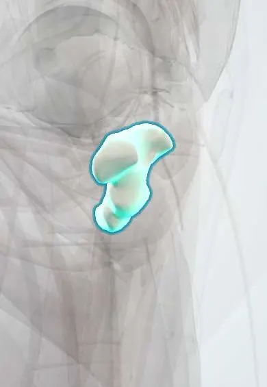
Coccyx !
The coccyx, or tailbone, is the small bone at the very bottom of the spine, just below the sacrum.
Opens in the Durgical app, no sign-up needed.
What it is
The tailbone: three to five small fused bones at the very bottom of the spine.
What it does
Anchors ligaments and pelvic floor muscles, and takes some weight when you lean back while sitting.
Key facts
- Joints with
- Sacrum (sacrococcygeal joint)
- Landmarks
- 3–5 fused rudimentary vertebrae (usually 4); coccygeal cornua; attachment of gluteus maximus, coccygeus, levator ani and anococcygeal ligament
Relations
- In front
- Ganglion impar; the rectum and anococcygeal body below and in front
- Behind
- Skin and fat of the natal cleft; dorsal sacrococcygeal ligaments
- Lateral
- Coccygeus and the iliococcygeus part of levator ani; edge of the sacrotuberous and sacrospinous ligaments
- Above
- Sacrum at the sacrococcygeal joint, a symphysis that may fuse with age
- Below
- Tip with the anococcygeal ligament running to the external anal sphincter
How to find it
Felt in the natal cleft a few centimetres behind the anus as the lower continuation of the sacrum. On rectal examination it can be held between a finger inside and the thumb outside to judge its tenderness and mobility.
Examination
- Rectal manipulation: moving the coccyx between a gloved finger in the rectum and the thumb outside reproduces the usual pain in coccydynia and shows whether the bone is too mobile or fixed
On imaging
A lateral view shows its angle; lateral views taken standing and sitting can show abnormal mobility (hypermobility or subluxation) in coccydynia. Normal angulation and segmentation vary a lot and are easily mistaken for fractures.
Common problems
- Coccydynia: tailbone pain, often after a fall or childbirth.
Clinical notes
- A fall onto the buttocks can fracture or dislocate the sacrococcygeal joint, sometimes with the coccyx displaced forward on rectal examination
- Coccydynia is more common in women; childbirth and rapid weight loss are recognised triggers
Anatomical variants
- Its shape ranges from a gentle forward curve to a sharp forward angle or a backward tilt; some shapes are linked to coccydynia
Development
The remnant of the embryonic tail, which regresses by about the eighth week of development.
Related conditions
Test yourself
Think of the answer, then tap the question to see it.
Which sympathetic structure lies in front of the sacrococcygeal junction and is blocked for coccygeal and perineal pain?
The ganglion impar, where the two sympathetic trunks end.
What is the commonest tumour in newborns related to the coccyx, and where does it come from?
Sacrococcygeal teratoma, from remnants of the primitive streak.
What are recognised triggers of coccydynia?
A fall onto the buttocks, childbirth and rapid weight loss; it is more common in women.
Educational information, not medical advice. For any symptom, see a health professional.
Its names
- Official Latin (TA2)
- os coccygis TA2 1092
- Official Spanish (DeCS)
- Cóccix DeCS D003050
- English (TA2)
- Coccyx
- Also called
- coccygeal bone · vertebrae coccygeae · vertebrae coccygeae I-IV · vertebrae Co1-Co4 · vertebrae coccygicae · vertebrae caudales
Related
Sources: Terminologia Anatomica (TA2); DeCS; Anatomy, Back, Coccygeal Vertebrae.
Spotted a mistake?We are always improving for the community. Tell us what is wrong and our team will review it.Report a mistake
Representative model, for educational purposes only.