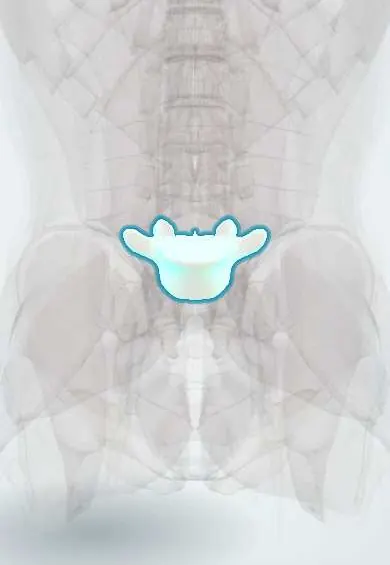
L5 vertebra !
L5 is the lowest lumbar vertebra, wedge-shaped, sitting on the sacrum; it passes the weight of the upper body to the pelvis.
Official name: L5
Opens in the Durgical app, no sign-up needed.
What it is
The lowest lumbar vertebra, wedge-shaped, sitting on the sacrum at the base of the spine.
What it does
Passes the weight of the whole upper body onto the sacrum and pelvis.
Key facts
- Joints with
- L4 above (disc, facet joints); sacrum (S1) below (L5–S1 disc and facet joints); tied to ilium by iliolumbar ligament
- Landmarks
- Largest body, wedge-shaped (taller in front), forming the lumbosacral angle; thick conical transverse processes; smallest lumbar spine; commonest site of spondylolysis
Relations
- In front
- The left common iliac vein crosses in front of L5 under the right common iliac artery; the superior hypogastric plexus and median sacral artery lie on the front of the L5–S1 disc
- Lateral
- The iliolumbar ligament runs from the thick transverse process to the iliac crest; the L5 ventral ramus joins L4 to form the lumbosacral trunk across the sacral ala
- Behind
- Low-back facet joints set more coronally than above, resisting forward shear on the sloping sacrum; the pars interarticularis between upper and lower facets
- Below
- Sacrum: the L5–S1 disc on the sacral promontory, the most wedge-shaped disc
How to find it
The L5 spine is the last lumbar spine felt in the midline, a little below the line across the iliac crests, just above the sacrum. The skin dimples over the posterior superior iliac spines lie lower, at about S2.
Examination
- One-leg hyperextension (stork) test: standing on one leg and arching back reproduces pain from a pars stress fracture, usually on the standing side
On imaging
Oblique views show the Scottie dog; a lateral view grades the slip and the slip angle. CT shows the pars gap; MRI shows an early stress reaction before a gap appears. On the AP view an enlarged L5 transverse process meeting the sacrum marks a transitional vertebra (Castellvi types).
Common problems
- Spondylolysis: a stress fracture of the bony bridge at the back (pars), common in young athletes who arch the back (gymnasts, football linemen).
- Isthmic spondylolisthesis: L5 slips forward on the sacrum after a pars fracture.
- L5-S1 disc herniation pressing on the S1 root.
How it is diagnosed
X-rays (including oblique and flexion-extension views) show slipping; CT shows a pars fracture; MRI shows discs and nerves.
Treatment
- Rest from the sport, physical therapy and sometimes a brace for a new pars fracture.
- Disabling slip or nerve pain: decompression and fusion at L5-S1 (posterior or anterior).
Clinical notes
- Bertolotti syndrome: low back pain linked to an enlarged L5 transverse process that forms a false joint with the sacrum or ilium
- Anterior approach to L5–S1 works between the common iliac vessels and can injure the superior hypogastric plexus, causing retrograde ejaculation in men
- May–Thurner syndrome: the left common iliac vein is squeezed between the right common iliac artery and L5, a cause of left leg deep vein thrombosis
Anatomical variants
- Sacralization of L5 or lumbarization of S1 (lumbosacral transitional vertebrae) are common, with frequencies that vary widely between studies
- Spina bifida occulta of L5 or S1: a gap in the lamina, usually without symptoms; a hairy patch or dimple above may mark a deeper defect
Development
Spondylolysis is acquired, not congenital: it is not seen in newborns and appears with upright activity in childhood and adolescence, more often in sports with repeated arching of the back.
Memory aid
Scottie dog: nose = transverse process, eye = pedicle, ear = superior articular process, neck = pars interarticularis, front leg = inferior articular process; a collar on the neck is a pars fracture.
Related conditions
Test yourself
Think of the answer, then tap the question to see it.
In isthmic spondylolisthesis at L5–S1, which nerve root is trapped, compared with an L5–S1 disc herniation?
The exiting L5 nerve root is trapped in the foramen; an L5–S1 disc herniation usually hits the traversing S1 nerve root.
On an oblique X-ray of the L5 vertebra, what does a collar on the Scottie dog's neck mean?
Spondylolysis: the neck is the pars interarticularis, and a dark collar across it is a pars fracture.
How does the Meyerding system grade a slip of L5 on S1?
In quarters of the S1 width: I up to 25%, II 25–50%, III 50–75%, IV 75–100%, V complete slip (spondyloptosis).
What is May–Thurner syndrome, which involves the L5 vertebra?
The left common iliac vein is squeezed between the right common iliac artery and L5, a cause of left leg deep vein thrombosis.
What is Bertolotti syndrome?
Low back pain linked to an enlarged L5 transverse process that forms a false joint with the sacrum or ilium.
Educational information, not medical advice. For any symptom, see a health professional.
Its names
- Official Latin (TA2)
- vertebra lumbalis V TA2 1068
- Official Spanish (DeCS)
- Vértebra lumbar L5 DeCS D008159
- English (TA2)
- L5
Related
- L4–L5 disc
- L5–S1 disc
- Iliolumbar ligament
- Iliolumbar vessels
- Anterior longitudinal ligament
- Endplates
- Low-back facet joints
- Interspinales
Sources: Terminologia Anatomica (TA2); DeCS; Anatomy, Back, Lumbar Spine.
Spotted a mistake?We are always improving for the community. Tell us what is wrong and our team will review it.Report a mistake
Representative model, for educational purposes only.