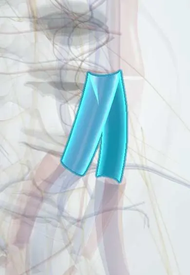
Common iliac veins !
The common iliac veins are two large veins that join in front of L5 to form the inferior vena cava; they drain the legs and pelvis.
Official name: Common iliac vein
Opens in the Durgical app, no sign-up needed.
What it is
The two large veins that join in front of the L5 vertebra (just right of centre) to form the inferior vena cava.
What it does
Return blood from the legs and pelvis. The left one crosses over L4-L5 and L5-S1 and is the main thing to protect in ALIF.
Key facts
- Drains
- Internal and external iliac veins (joining in front of the sacroiliac joint); iliolumbar veins; left also receives the median sacral vein
- Into
- Inferior vena cava at L5, right of the midline; left vein crossed by the right common iliac artery (May-Thurner compression)
Relations
- Course
- Each forms in front of the sacroiliac joint from the external and internal iliac veins; the right rises almost vertically behind its artery, the left runs obliquely across the front of L5, and they meet at L5 just right of the midline to form the inferior vena cava
- In front
- The right common iliac artery, which crosses the left vein at L5, and the peritoneum
- Behind
- L5 vertebral body, the L4–L5 and L5–S1 discs, the sacral promontory and the lumbosacral trunk; the iliolumbar and ascending lumbar veins enter from behind and tether the left vein
- Medial
- The median sacral vein, which usually drains into the left common iliac vein, and the L5–S1 disc between the two veins
How to find it
They cannot be felt. In anterior exposure they join to form the inferior vena cava at about L5, below and to the right of the aortic bifurcation; the left vein crosses the L5 body obliquely, so L5–S1 is usually reached between the iliac vessels and L4–L5 by moving the left iliac vessels to the right.
On imaging
CT or MR venography and intravascular ultrasound show narrowing of the left vein where the right artery crosses it, often with collateral veins across the pelvis.
Clinical notes
- To expose L4–L5 from the front, the left common iliac vein is moved to the right only after the iliolumbar and ascending lumbar veins that tether it have been tied
- A tear in its thin wall bleeds briskly; it is usually controlled with gentle pressure from small sponge sticks and then sutured
- May-Thurner syndrome is treated endovascularly in selected cases, with clot removal and a stent at the point of compression
- Thrombus can extend from the iliac veins into the inferior vena cava and give rise to pulmonary embolism
Anatomical variants
- The way the internal and external iliac veins join varies, for example a double internal iliac vein or a higher or lower confluence
- With a left-sided or duplicated inferior vena cava the left common iliac vein drains differently, which changes the anterior approach
Development
The left common iliac vein starts as a cross channel that carries blood from the left leg over to the right-sided venous trunk that becomes the inferior vena cava; this is why it is longer and crosses the midline.
Test yourself
Think of the answer, then tap the question to see it.
Where do the common iliac veins join to form the inferior vena cava?
At L5, just right of the midline.
What is May-Thurner syndrome?
Compression of the left common iliac vein between the right common iliac artery and L5, predisposing to left leg swelling and deep vein thrombosis.
Which vessel is most often torn in anterior lumbar spine surgery?
The left common iliac vein; venous injury is more common than arterial injury in these approaches.
Which veins must be tied before the left common iliac vein is moved to expose L4–L5 from the front?
The iliolumbar and ascending lumbar veins, which tether it.
Educational information, not medical advice. For any symptom, see a health professional.
Its names
- Official Latin (TA2)
- vena iliaca communis TA2 5021
- Official Spanish (DeCS)
- Venas ilíacas comunes DeCS D007084
- English (TA2)
- Common iliac vein
- Also called
- vena ilica communis
Related
Sources: Terminologia Anatomica (TA2); DeCS; Anatomy, Abdomen and Pelvis: Veins; Anatomy of the Human Body (20th ed.): Development of the Vascular System (Lea & Febiger (Gray's Anatomy, 1918), 1918) · adapted, Public domain.
Spotted a mistake?We are always improving for the community. Tell us what is wrong and our team will review it.Report a mistake
Representative model, for educational purposes only.