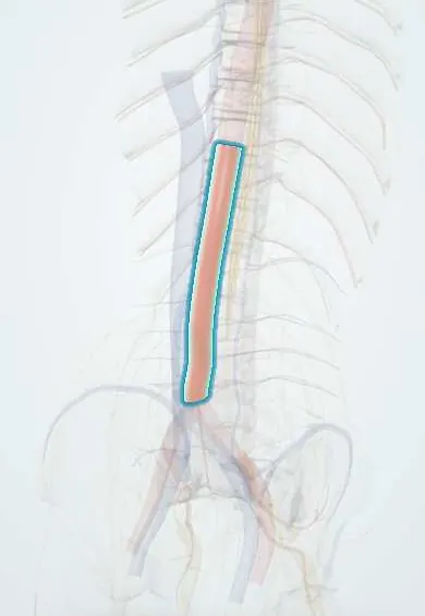
Abdominal aorta !
The abdominal aorta is the body's main artery in the abdomen, lying on the front left of the lumbar spine; it splits at about L4.
Opens in the Durgical app, no sign-up needed.
What it is
The body's main artery in the abdomen, lying on the front left of the lumbar vertebrae and splitting into the two common iliac arteries at about L4.
What it does
Carries blood from the heart to the abdomen, pelvis and legs, and gives off the lumbar arteries that feed the spine.
Key facts
- Origin
- Continuation of thoracic aorta at the aortic hiatus (T12); bifurcates at L4 into the common iliac arteries
- Branches
- Celiac trunk (T12), superior mesenteric (L1), renal (L1–L2), inferior mesenteric (L3); inferior phrenic, suprarenal, gonadal; 4 pairs of lumbar; median sacral
- Supplies
- Abdominal viscera, posterior abdominal wall, lumbar spine and cauda equina, pelvis and lower limbs
Relations
- Course
- From the aortic hiatus (T12) it runs down on the front of the L1–L4 vertebral bodies, slightly left of the midline, to its bifurcation at L4
- In front
- Coeliac plexus, body of the pancreas and splenic vein, left renal vein, third part of the duodenum and the root of the mesentery, all under the peritoneum
- Behind
- Anterior longitudinal ligament and the L1–L4 vertebral bodies; the left lumbar veins cross behind it to reach the inferior vena cava
- Lateral
- Right: the inferior vena cava, the cisterna chyli and, above, the right crus of the diaphragm; left: the left sympathetic trunk, the left psoas major and the duodenojejunal flexure
How to find it
Lying supine with the knees bent and the abdomen relaxed, it is felt by deep pressure with both hands just above and slightly left of the umbilicus. A normal aorta pulses forward; a pulsation that pushes the hands apart (expansile) suggests an aneurysm. In thin people a visible pulsation can be normal.
On imaging
Ultrasound in transverse and longitudinal planes, from the xiphoid to the bifurcation, measures the maximum diameter; CT angiography maps the neck, the branches and the iliac arteries before repair. A calcified wall is often seen in front of the lumbar bodies on a lateral radiograph.
Common problems
- An abdominal aortic aneurysm (a ballooning of the artery) can cause back pain and must not be mistaken for a spine problem.
- Its position and calcium matter when planning anterior lumbar surgery.
How it is diagnosed
Ultrasound or CT angiography.
Treatment
- Aneurysms are treated by vascular surgeons. In ALIF, an access surgeon gently moves the vessels aside to reach the disc.
Clinical notes
- Screening programmes offer a one-time abdominal ultrasound to older men who have ever smoked (for example aged 65–75), because rupture is often fatal
- A ruptured or leaking aneurysm classically gives back or flank pain, low blood pressure and a pulsatile mass, and can be mistaken for renal colic
- Aortic dissection can extend into it and cut off the lumbar arteries or the visceral branches, causing spinal cord or bowel ischaemia
- It lies just in front of the upper lumbar discs and, with the iliac arteries, the L4–L5 disc: an instrument pushed through the anterior annulus in posterior disc surgery can tear a great vessel, a rare but life-threatening complication
- Leriche syndrome: aortoiliac occlusion with buttock and thigh claudication, weak or absent femoral pulses and erectile dysfunction
Anatomical variants
- Accessory renal arteries are common, seen in about a quarter of people
- The bifurcation is usually at L4 but ranges from about L3 to L5
- The coeliac trunk and superior mesenteric artery occasionally arise from a common trunk
Development
The paired dorsal aortae of the embryo merge into one vessel from about the fourth thoracic to the fourth lumbar segment; their segmental branches persist as the lumbar arteries, and the thin tail end persists as the median sacral artery.
Memory aid
Openings of the diaphragm: "I 8 10 Eggs At 12": Inferior vena cava at T8, oEsophagus at T10, Aorta at T12.
Related conditions
Test yourself
Think of the answer, then tap the question to see it.
At which vertebral levels does the abdominal aorta begin and bifurcate?
It begins at the aortic hiatus at T12 and divides into the common iliac arteries at L4, roughly level with the iliac crests.
What diameter defines an abdominal aortic aneurysm, and where do most begin?
A diameter of 3 cm or more; most begin below the renal arteries (infrarenal).
What is the classic presentation of a ruptured abdominal aortic aneurysm?
Back or flank pain, low blood pressure and a pulsatile mass; it can be mistaken for renal colic.
Which vein crosses in front of the abdominal aorta, and what is its compression called?
The left renal vein, just below the superior mesenteric artery; compression in that angle is the nutcracker phenomenon.
What is Leriche syndrome?
Aortoiliac occlusion with buttock and thigh claudication, weak or absent femoral pulses and erectile dysfunction.
Educational information, not medical advice. For any symptom, see a health professional.
Its names
- Official Latin (TA2)
- aorta abdominalis TA2 4205
- Official Spanish (DeCS)
- Aorta abdominal DeCS D001012
- English (TA2)
- Abdominal aorta
- Also called
- Aorta, Abdominal
Related
Sources: Terminologia Anatomica (TA2); DeCS; Anatomy, Abdomen and Pelvis: Abdominal Aorta; Anatomy of the Human Body (20th ed.): Development of the Vascular System (Lea & Febiger (Gray's Anatomy, 1918), 1918) · adapted, Public domain.
Spotted a mistake?We are always improving for the community. Tell us what is wrong and our team will review it.Report a mistake
Representative model, for educational purposes only.