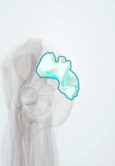
Sacrum !
The sacrum is the triangle-shaped bone at the base of the spine; it joins the two hip bones to close the back of the pelvis.
Opens in the Durgical app, no sign-up needed.
What it is
A large, triangle-shaped bone made of five fused vertebrae at the base of the spine, wedged between the two hip bones.
What it does
Passes the weight of the upper body to the pelvis through the sacroiliac joints.
Key facts
- Joints with
- L5 (lumbosacral joint); coccyx; both hip bones (sacroiliac joints)
- Landmarks
- Five fused vertebrae; promontory; alae; four pairs of anterior and posterior sacral foramina; median sacral crest; sacral hiatus; auricular surface
Relations
- Above
- Base with L5 at the lumbosacral disc; the lumbosacral trunk (L4–L5) crosses the ala
- In front
- Sacral plexus roots from the anterior foramina, piriformis origin, sympathetic trunks and median sacral vessels; the rectum and mesorectum in front
- Behind
- Median and lateral sacral crests; erector spinae and multifidus; part of the origin of gluteus maximus
- Lateral
- Auricular surface forming the sacroiliac joint with the ilium; the sacrotuberous and sacrospinous ligaments on its lateral border
- Below
- Apex with the coccyx at the sacrococcygeal joint
- Contents
- Sacral canal: cauda equina roots, the lower end of the dural sac and the filum terminale
How to find it
The skin dimples over the posterior superior iliac spines mark S2 and the middle of the sacroiliac joints. The median sacral crest is felt above the natal cleft; the sacral hiatus is the hollow between the two cornua at the top of the cleft, just above the coccyx. The front is felt on rectal examination; the promontory, upper end of the diagonal conjugate, may be reached vaginally.
Examination
- Sacroiliac provocation tests (distraction, thigh thrust, compression, sacral thrust, Gaenslen): three or more positive out of five support the sacroiliac joint as the source of pain
- FABER (Patrick) test: with the hip flexed, abducted and externally rotated, pain over the back of the sacroiliac joint points to a sacroiliac source, while groin pain points to the hip
On imaging
Easy to miss on the AP pelvis because of its tilt and overlying bowel gas: the arcuate lines of the sacral foramina are checked for breaks. CT for fractures; MRI or bone scan for insufficiency fractures. Lumbosacral transitional vertebrae are counted from the top of the spine to avoid level errors.
Common problems
- Stress fractures (in older adults with weak bones, or in runners) and fractures from falls or accidents.
Clinical notes
- Caudal epidural block through the sacral hiatus is widely used in children
- The sacroiliac joint is a source of low back and buttock pain; sacroiliitis is an early sign of ankylosing spondylitis
- A fall onto the buttocks or a high-energy pelvic injury can fracture it; a transverse fracture with kyphosis threatens the sacral roots
Anatomical variants
- Lumbosacral transitional vertebra (sacralised L5 or lumbarised S1), up to about 12% in a meta-analysis; it changes level counting before surgery or injections
- Sacral spina bifida occulta is common and usually harmless; the number of foramina varies
- The female sacrum is shorter and wider, with a less prominent promontory
Development
Five sacral vertebrae, each with a body, arches and lateral (costal) elements that build the alae, fuse progressively from adolescence until about the mid-twenties.
Related conditions
- Spondylolisthesis
- Sacroiliac joint pain
- Spina bifida (myelomeningocele)
- Pressure injury (pressure ulcer)
- Immobility syndrome and hospital-associated deconditioning
- Incontinence-associated dermatitis
- Pelvic fracture
- Pilonidal disease
Test yourself
Think of the answer, then tap the question to see it.
What is the entry point for a caudal epidural block on the sacrum?
The sacral hiatus, between the sacral cornua where the S5 laminae are missing.
At what level does the dural sac usually end, and what surface landmark marks it?
At S2, level with the posterior superior iliac spines (the skin dimples).
What is the Honda sign of the sacrum on bone scan?
H-shaped uptake from a sacral insufficiency fracture, typical of older adults with osteoporosis.
Which sacral fractures put the sacral nerve roots at risk?
Fractures through the foramina or the central canal (Denis zones II and III), with bladder and bowel effects.
Educational information, not medical advice. For any symptom, see a health professional.
Its names
- Official Latin (TA2)
- os sacrum TA2 1071
- Official Spanish (DeCS)
- Sacro DeCS D012447
- English (TA2)
- Sacrum
- Also called
- sacral bone · vertebrae sacrales · vertebrae sacrales I-V · vertebrae S1-S5
Related
- Sacroiliac ligaments
- Coccyx
- Sacrotuberous ligament
- Sacrospinous ligament
- Fascia lata
- Gluteus maximus
- Iliopsoas fascia
- Piriformis
Sources: Terminologia Anatomica (TA2); DeCS; Anatomy, Back, Sacral Vertebrae; Anatomía humana (4.ª ed.), tomo 1, cap. 4: Esqueleto de la columna vertebral.
Spotted a mistake?We are always improving for the community. Tell us what is wrong and our team will review it.Report a mistake
Representative model, for educational purposes only.