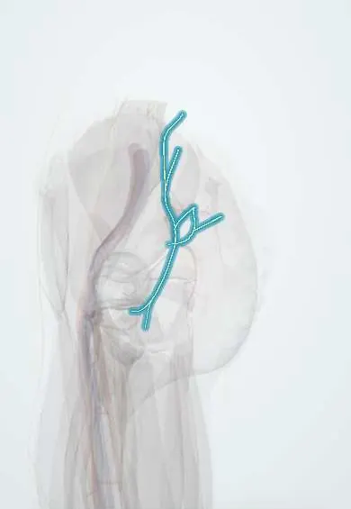
Nerves to the hip rotators !
The nerves to the hip rotators are three small nerves from the sacral plexus supplying the deep buttock muscles that turn the thigh out.
Official name: Nerve to piriformis muscle
Opens in the Durgical app, no sign-up needed.
What it is
Small nerves from the sacral plexus: the nerve to the piriformis and the nerve to the quadratus femoris.
What it does
Control deep muscles that turn the thigh outward (piriformis, quadratus femoris and inferior gemellus) and send small branches to the hip joint.
Key facts
- Roots
- Nerve to piriformis S1–S2; nerve to quadratus femoris L4–S1; nerve to obturator internus L5–S2
- Moves
- Piriformis; quadratus femoris and inferior gemellus; obturator internus and superior gemellus
- Feeling from
- None to skin; nerve to quadratus femoris gives twigs to hip joint
Relations
- Course
- The nerve to the piriformis stays in the pelvis and enters the muscle's pelvic surface; the other two leave below the piriformis; the nerve to the obturator internus then curves around the ischial spine into the lesser sciatic foramen, and the nerve to the quadratus femoris descends on the ischium
- Medial
- The internal pudendal vessels and, most medial, the pudendal nerve, which cross the ischial spine beside the nerve to the obturator internus
- Superficial
- The sciatic nerve, and the gemelli and obturator internus tendon, cover the nerve to the quadratus femoris
- Deep
- The posterior surface of the ischium and the back of the hip joint capsule
How to find it
They cannot be felt. In dissection, with the gluteus maximus lifted, the nerve to the obturator internus is found medial to the sciatic nerve near the ischial spine; the nerve to the quadratus femoris appears only when the gemelli and obturator internus tendon are turned aside.
On imaging
Usually not seen on their own. They are assessed through the muscles they supply: on MRI, denervation oedema or fatty atrophy in the piriformis, obturator internus, gemelli or quadratus femoris points to a lesion. Their landmarks are the sciatic nerve, which they run beside, and the ischial spine. MR neurography may show the larger ones. Radiographs and CT do not show them.
Clinical notes
- In the posterior hip approach the short external rotators are detached near the femur and folded back over the sciatic nerve to protect it
- The nerve to the obturator internus is a landmark in the buttock for the pudendal nerve, which lies just medial to it at the ischial spine
Memory aid
Over the ischial spine, medial to lateral: 'PIN' (Pudendal nerve, Internal pudendal vessels, Nerve to obturator internus).
Test yourself
Think of the answer, then tap the question to see it.
Which three small sacral plexus nerves supply the short hip rotators?
The nerve to piriformis, the nerve to obturator internus (with superior gemellus) and the nerve to quadratus femoris (with inferior gemellus).
What path does the nerve to obturator internus take at the ischial spine?
It leaves through the greater sciatic foramen and re-enters through the lesser, crossing the ischial spine beside the pudendal nerve.
Which of the nerves to the hip rotators sends twigs to the hip joint?
The nerve to quadratus femoris, which lies deep to the gemelli and obturator internus tendon on the back of the capsule.
Educational information, not medical advice. For any symptom, see a health professional.
Its names
- Official Latin (TA2)
- nervus musculi piriformis; nervus obturatoris interni; nervus musculi quadrati femoris TA2 6545
- Official Spanish (DeCS)
- Nervios del piriforme, del obturador interno y del cuadrado femoral
- English (TA2)
- Nerve to piriformis muscle
- Also called
- nerve to obturator internus muscle
Related
Sources: Terminologia Anatomica (TA2); Anatomy, Bony Pelvis and Lower Limb: Nerves.
Spotted a mistake?We are always improving for the community. Tell us what is wrong and our team will review it.Report a mistake
Representative model, for educational purposes only.