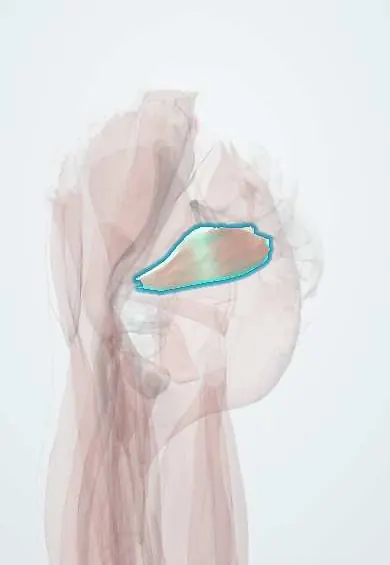
Piriformis !
The piriformis is a deep buttock muscle running from the sacrum to the femur; it turns the thigh outward and lies near the sciatic nerve.
Official name: Piriformis muscle
Opens in the Durgical app, no sign-up needed.
What it is
A pear-shaped muscle deep in the buttock, from the front of the sacrum, out through the greater sciatic notch, to the top of the greater trochanter. The sciatic nerve usually passes just below it.
What it does
Turns the thigh outward when the hip is straight, moves it out to the side when the hip is bent, and helps hold the ball in the socket.
Key facts
- Origin
- Anterior surface of sacrum (S2–S4 level); sacrotuberous ligament; leaves pelvis through greater sciatic foramen
- Insertion
- Superior border of greater trochanter
- Action
- Laterally rotates extended hip; abducts flexed hip; steadies femoral head
- Nerve
- Nerve to piriformis (S1–S2) (some texts: L5–S2)
- Blood supply
- Superior and inferior gluteal arteries
Relations
- Course
- From the front of the sacrum inside the pelvis it passes out through the greater sciatic foramen, nearly filling it, and runs laterally behind the hip joint to the top of the greater trochanter
- In front
- Inside the pelvis, the sacral plexus lies on its front surface, with the rectum medially
- Above
- Gluteus medius and gluteus minimus; the superior gluteal vessels and nerve emerge along its upper border
- Below
- Superior gemellus; the sciatic nerve and the other infrapiriform structures emerge along its lower border
- Superficial
- Gluteus maximus
How to find it
It cannot be felt directly. Its line runs from the lower sacrum, about midway between the posterior superior iliac spine and the coccyx, to the top of the greater trochanter; deep tenderness along this line is sought with the hip flexed, adducted and turned inward. Ultrasound, CT or MRI guides injections.
Examination
- FAIR test: lying on the unaffected side, the hip is flexed, adducted and turned inward; buttock or leg pain suggests the muscle is irritating the sciatic nerve. No test is specific, so a lumbar radiculopathy is excluded first
- Freiberg sign: pain on passive internal rotation of the straight hip
- Pace sign: pain or weakness when the seated person pushes the knees apart (abduction and external rotation) against resistance
On imaging
MRI is best: on axial and coronal images it runs from the front of the sacrum through the greater sciatic foramen to the greater trochanter, with the sciatic nerve below it. Look for asymmetry, oedema or a nerve passing through the muscle. Imaging is often normal in suspected piriformis syndrome, which stays a clinical diagnosis. Ultrasound or CT guides injections.
Common problems
- Piriformis syndrome: deep buttock pain, sometimes with sciatica-like pain down the leg, thought to come from the muscle irritating the sciatic nerve.
- Pain with long sitting.
- In some people the nerve passes through or above the muscle, a normal variation.
How it is diagnosed
Mostly by exam, after ruling out other causes such as a spinal disc problem. MRI of the spine and pelvis; sometimes a diagnostic injection.
Treatment
- Physical therapy with stretching and strengthening; avoiding long sitting.
- Ultrasound-guided injection into the muscle.
- Rarely, surgery to release the muscle when other treatment fails.
Clinical notes
- Posterior approach to the hip: its tendon and the short rotators are detached and later repaired; the sciatic nerve is found just below and behind it
- The classic posterior sciatic nerve block aims below the piriformis, using lines between the posterior superior iliac spine, the greater trochanter and the sacral hiatus
Anatomical variants
- Texts differ on frequency: the sciatic nerve or its common fibular part pierces the muscle or passes above it in roughly 1 in 10 to 1 in 5 people (cited review: at least 1 in 5); usually harmless, and a proposed factor in piriformis syndrome
- It may be fused with the gluteus medius or the gluteus minimus
Development
Develops from the dorsal muscle mass and is supplied by the nerve to piriformis (S1–S2), from posterior divisions of the sacral plexus; the short rotators below it belong to the ventral mass and take anterior-division nerves.
Memory aid
Structures below the piriformis: POP'S IQ (Pudendal nerve, nerve to Obturator internus, Posterior femoral cutaneous nerve, Sciatic nerve, Inferior gluteal nerve and vessels, nerve to Quadratus femoris).
Related conditions
Test yourself
Think of the answer, then tap the question to see it.
Which structures leave the greater sciatic foramen above the piriformis?
Only the superior gluteal nerve, artery and vein.
Which structures leave below the piriformis (POP'S IQ)?
Pudendal nerve, nerve to obturator internus, posterior femoral cutaneous nerve, sciatic nerve, inferior gluteal nerve and vessels, nerve to quadratus femoris.
What is the relevant sciatic nerve variant at the piriformis?
In a minority (roughly 1 in 10 to 1 in 5), the sciatic nerve or its common fibular part pierces the muscle or passes above it.
What is the FAIR test for piriformis syndrome?
Lying on the unaffected side, the hip is flexed, adducted and turned inward; buttock or leg pain suggests the piriformis is irritating the sciatic nerve.
What is the action of the piriformis?
It laterally rotates the extended hip, abducts the flexed hip and steadies the femoral head.
Educational information, not medical advice. For any symptom, see a health professional.
Its names
- Official Latin (TA2)
- musculus piriformis TA2 2604
- Official Spanish (DeCS)
- Músculo piriforme
- English (TA2)
- Piriformis muscle
Related
- Sciatic nerve
- Piriformis fascia
- Superior gemellus
- Gluteus medius
- Gluteal arteries
- Superior gluteal nerve
- Deep hip bursae
- Nerves to the hip rotators
Sources: Terminologia Anatomica (TA2); Anatomy, Bony Pelvis and Lower Limb: Piriformis Muscle.
Spotted a mistake?We are always improving for the community. Tell us what is wrong and our team will review it.Report a mistake
Representative model, for educational purposes only.