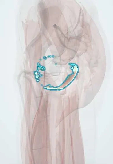
Obturator internus !
The obturator internus is a deep hip muscle that starts inside the pelvis and turns the thigh outward.
Opens in the Durgical app, no sign-up needed.
What it is
A fan-shaped muscle that starts inside the pelvis, around the obturator foramen. Its tendon bends around the hip bone and runs to the greater trochanter.
What it does
Turns the thigh outward and holds the ball steady in the socket.
Key facts
- Origin
- Pelvic surface of obturator membrane and surrounding bone; leaves pelvis through lesser sciatic foramen
- Insertion
- Medial surface of greater trochanter (with the gemelli)
- Action
- Laterally rotates extended hip; abducts flexed hip; steadies femoral head
- Nerve
- Nerve to obturator internus (L5–S1) (some texts: L5–S2)
- Blood supply
- Internal pudendal and obturator arteries
Relations
- Medial
- Obturator fascia and the origin of the levator ani; below the levator, the ischioanal fossa with the pudendal canal
- Course
- From the inner surface of the obturator membrane its fibres converge on a tendon that leaves the pelvis through the lesser sciatic foramen, turns sharply around the ischium and crosses the back of the hip joint to the greater trochanter
- Above
- Inside the pelvis, the obturator canal with the obturator nerve and vessels; in the gluteal region, the superior gemellus
- Below
- Inferior gemellus, then quadratus femoris
- Superficial
- Sciatic nerve, which crosses its tendon and the gemelli under the gluteus maximus
How to find it
Not palpable from outside. On rectal or vaginal examination it can be felt on the side wall of the pelvis above the levator ani, tightening when the thigh is turned outward against resistance.
Examination
- Obturator sign: with the hip and knee bent to 90°, the hip is rotated inward; lower abdominal pain suggests an inflamed pelvic appendix or an abscess irritating the muscle
On imaging
MRI and CT show it lining the side wall of the lesser pelvis, with its tendon turning round the lesser sciatic notch to the greater trochanter. On MRI look for strain, oedema or an abscess, often next to a pelvic infection. Its fascia forms the lateral wall of the ischioanal fossa, where perianal abscesses spread. Ultrasound or CT guides injections. Radiographs do not show it.
Common problems
- Uncommonly, a source of deep buttock or pelvic pain when strained or tight.
Clinical notes
- Pudendal nerve block and pudendal nerve entrapment (for example in cyclists) involve the pudendal canal in its fascia and the nearby ischial spine
- In the posterior hip approach, the short rotators are detached and folded back over the sciatic nerve to protect it, then repaired
Anatomical variants
- Its tendon joins the piriformis tendon before inserting in about 1 in 10 hips in the cited review
Development
A ventral-mass muscle: its nerve (L5–S2) comes from anterior divisions of the sacral plexus, unlike that of the piriformis just above, which belongs to the dorsal mass.
Related conditions
Test yourself
Think of the answer, then tap the question to see it.
Through which foramen does the obturator internus tendon leave the pelvis?
The lesser sciatic foramen, turning almost at a right angle around the lesser sciatic notch, which acts as a pulley.
Which canal is formed by the obturator fascia over the obturator internus, and what does it carry?
The pudendal (Alcock) canal, carrying the pudendal nerve and internal pudendal vessels.
What does the obturator sign suggest?
With hip and knee at 90°, internal rotation causing lower abdominal pain suggests an inflamed pelvic appendix or abscess irritating the obturator internus.
What is the nerve supply of the obturator internus?
The nerve to obturator internus (L5–S1).
Educational information, not medical advice. For any symptom, see a health professional.
Its names
- Official Latin (TA2)
- obturator internus TA2 2605
- Official Spanish (DeCS)
- Músculo obturador interno
- English (TA2)
- Obturator internus
- Also called
- obturator internus muscle
Related
- Obturator membrane
- Superior gemellus
- Inferior gemellus
- Gluteal arteries
- Obturator artery
- Obturator internus tendon
- Femur
- Hip bone
Sources: Terminologia Anatomica (TA2); Anatomy, Abdomen and Pelvis, Obturator Muscles.
Spotted a mistake?We are always improving for the community. Tell us what is wrong and our team will review it.Report a mistake
Representative model, for educational purposes only.