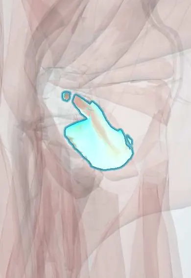
Obturator externus !
The obturator externus is a deep muscle that wraps under and behind the hip joint from the pelvis; it turns the thigh outward.
Opens in the Durgical app, no sign-up needed.
What it is
A flat, fan-shaped muscle on the outer side of the obturator membrane. Its tendon winds under and behind the neck of the femur.
What it does
Turns the thigh outward and steadies the ball in the socket.
Key facts
- Origin
- External surface of obturator membrane and margins of obturator foramen
- Insertion
- Trochanteric fossa of femur
- Action
- Laterally rotates hip; steadies femoral head in acetabulum
- Nerve
- Obturator nerve, posterior branch (L3–L4) (some texts: L2–L4)
- Blood supply
- Obturator artery; medial circumflex femoral artery
Relations
- In front
- Pectineus and adductor brevis
- Behind
- Quadratus femoris, with the deep branch of the medial circumflex femoral artery crossing behind its tendon
- Above
- Obturator canal: the obturator nerve and vessels emerge above it
- Deep
- Obturator membrane, with the obturator internus on its pelvic side
- Course
- Its tendon passes below the femoral neck and behind the hip joint capsule to the trochanteric fossa
How to find it
Not palpable. MRI shows it in the upper medial thigh deep to the pectineus and the adductor brevis, with its tendon passing under and behind the femoral neck on axial and coronal images.
Examination
- Howship–Romberg sign: pain along the inner thigh toward the knee, worse with hip extension, abduction and internal rotation, from an obturator hernia pressing on the obturator nerve
On imaging
MRI shows it well: on coronal images it spreads from the outer surface of the obturator membrane and winds below and behind the femoral neck to the trochanteric fossa. Oedema or a strain appears in athletes with groin pain. It is checked in hip fractures and dislocations because of its link with the medial circumflex vessels behind its tendon. Radiographs do not show it.
Clinical notes
- An obturator hernia passes through the obturator canal above the muscle and can lie between it and the pectineus; it is rare, seen mostly in thin older women, and often found late
- Surgical dislocation of the hip (Ganz) keeps this muscle intact to protect the blood supply of the femoral head
- Strains occur in sports with forceful hip rotation and appear on MRI as oedema in the muscle; they can mimic other causes of deep groin or hip pain
Development
From the ventral muscle mass, like the adductors; this is why it shares their obturator nerve (anterior divisions of L2–L4) although it works as a lateral rotator.
Test yourself
Think of the answer, then tap the question to see it.
Which nerve supplies the obturator externus, and what is its action?
The obturator nerve, posterior branch (L3–L4), like the adductors, but it works as a lateral rotator of the hip.
Why is the obturator externus tendon kept intact in surgical dislocation of the hip (Ganz)?
The deep branch of the medial circumflex femoral artery, the main supply of the femoral head, runs just behind it.
How do the branches of the obturator nerve relate to the obturator externus?
The nerve divides at its upper border: the anterior branch passes above the muscle, the posterior branch pierces it.
Educational information, not medical advice. For any symptom, see a health professional.
Its names
- Official Latin (TA2)
- obturator externus TA2 2636
- Official Spanish (DeCS)
- Músculo obturador externo
- English (TA2)
- Obturator externus
- Also called
- obturator externus muscle
Related
- Obturator membrane
- Quadratus femoris
- Obturator nerve
- Circumflex femoral arteries
- Obturator artery
- Obturator externus tendon
- Femur
- Hip bone
Sources: Terminologia Anatomica (TA2); Anatomy, Abdomen and Pelvis, Obturator Muscles; Anatomy, Bony Pelvis and Lower Limb: Medial Thigh Muscles.
Spotted a mistake?We are always improving for the community. Tell us what is wrong and our team will review it.Report a mistake
Representative model, for educational purposes only.