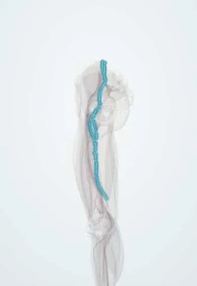
Obturator nerve !
The obturator nerve runs from the pelvis into the inner thigh; it controls the muscles that pull the leg inward.
Opens in the Durgical app, no sign-up needed.
What it is
A nerve from the lower spine that runs along the inside wall of the pelvis and exits through the obturator canal into the inner thigh, where it splits into front and back branches.
What it does
Controls the adductor muscles that pull the leg inward, and carries feeling from a patch of skin on the inner thigh.
Key facts
- Roots
- L2–L4 (anterior divisions, lumbar plexus)
- Moves
- Adductor longus, brevis and adductor part of magnus, gracilis, obturator externus; sometimes pectineus
- Feeling from
- Variable patch of medial thigh; hip and knee joints (knee pain referred from hip)
Relations
- Course
- Leaves the medial border of the psoas major at the pelvic brim, passes behind the common iliac vessels, runs along the lateral pelvic wall on the obturator internus to the obturator canal and divides into an anterior division (in front of the adductor brevis) and a posterior division (behind it, through the obturator externus)
- Medial
- The ureter, the internal iliac vessels and their branches, the obturator lymph nodes and, in women, the ovary
- Lateral
- The obturator internus on the lateral pelvic wall
- Below
- The obturator artery and vein, which run just below it along the pelvic wall and through the canal
How to find it
It cannot be felt. Just below the pubis, ultrasound shows the anterior division between the adductor longus and adductor brevis and the posterior division between the adductor brevis and adductor magnus, where it is blocked. At laparoscopy it is seen on the lateral pelvic wall below the external iliac vessels.
Examination
- Resisted hip adduction (squeezing a fist between the knees): weakness with normal knee extension points to the obturator nerve; quadriceps weakness as well points to the L3–L4 roots or the lumbar plexus
- Pinprick on the medial thigh: loss is inconstant because the cutaneous branch is small or absent in many people
On imaging
Ultrasound of the upper medial thigh shows the anterior branch between the adductor longus and brevis and the posterior branch between the brevis and magnus; this guides the obturator block. In the pelvis it lies on the side wall near the obturator canal on MRI. MR neurography or MRI looks for a compressing mass or for denervation oedema in the adductors. Radiographs do not show it.
Common problems
- Can be injured in pelvic surgery or childbirth. Hip joint pain can travel along it and be felt at the inner knee.
Clinical notes
- At risk in pelvic lymph node dissection for gynaecological and urological cancers, in acetabular fractures and screws, and in hip replacement
- Obturator hernia: typically a thin older woman with bowel obstruction and medial thigh pain down to the knee (Howship–Romberg sign)
- Obturator block prevents the sudden adductor jerk caused by electrical stimulation during transurethral resection of lateral bladder wall tumours, and is used for adductor spasticity
- Because it also supplies the hip and knee joints, hip disease (for example in children) may first appear as knee pain
Anatomical variants
- Where it divides varies: in the obturator canal in about half of people, otherwise in the thigh or in the pelvis (about a quarter each)
- An accessory obturator nerve that crosses over the superior pubic ramus behind the pectineus, instead of passing through the canal, is present in a minority of people
Development
Built from the anterior divisions of the ventral rami of L2–L4, it supplies muscles of the ventral muscle mass, which come to lie in the medial compartment of the thigh after the limb rotates.
Test yourself
Think of the answer, then tap the question to see it.
How does the obturator nerve reach the thigh?
It leaves the medial border of the psoas major, runs along the lateral pelvic wall and passes through the obturator canal.
Which muscles does the obturator nerve supply?
Adductor longus, adductor brevis, the adductor part of adductor magnus, gracilis and obturator externus; sometimes pectineus.
What gait results from an obturator nerve lesion?
A wide-based, outward-swinging gait from weak hip adduction.
Why may hip disease in a child first present as knee pain?
The obturator nerve supplies both the hip and knee joints, so hip pain can be referred to the knee.
Why is an obturator nerve block used during transurethral resection of lateral bladder wall tumours?
It prevents the sudden adductor jerk caused by electrical stimulation of the obturator nerve.
Educational information, not medical advice. For any symptom, see a health professional.
Its names
- Official Latin (TA2)
- nervus obturatorius TA2 6532
- Official Spanish (DeCS)
- Nervio obturador DeCS D009776
- English (TA2)
- Obturator nerve
Related
- Obturator membrane
- Obturator externus
- Adductor brevis
- Obturator artery
- Adductor longus
- Adductor magnus
- Adductor minimus
- Gracilis
Sources: Terminologia Anatomica (TA2); DeCS; Anatomy, Abdomen and Pelvis, Obturator Nerve; 8.5 Development of the Appendicular Skeleton (OpenStax (Rice University), 2022) · adapted, CC BY 4.0.
Spotted a mistake?We are always improving for the community. Tell us what is wrong and our team will review it.Report a mistake
Representative model, for educational purposes only.