Cardiovascular system · Lower limb
Obturator artery !
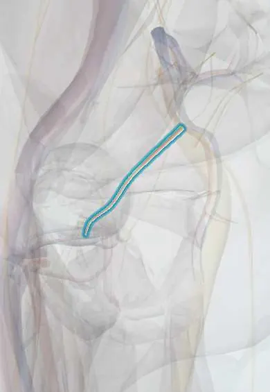
The obturator artery runs from the pelvis to the inner thigh; a small branch reaches the femoral head through its ligament.
Opens in the Durgical app, no sign-up needed.
What it is
A branch of the internal iliac artery that runs along the pelvic wall and through the obturator canal to the inner thigh.
What it does
Supplies the adductor and obturator muscles. A small branch runs in the ligament of the femoral head to the ball of the femur.
Key facts
- Origin
- Anterior division of internal iliac artery (varies: from inferior epigastric as aberrant obturator, about 20–30%)
- Branches
- Anterior and posterior branches; acetabular branch giving artery of ligament of femoral head
- Supplies
- Adductor muscles, obturator externus, hip joint; small supply to femoral head
Relations
- Course
- From the anterior division of the internal iliac artery it runs forward on the lateral pelvic wall below the obturator nerve, passes through the obturator canal and splits into anterior and posterior branches that ring the obturator foramen
- Above
- The obturator nerve
- Below
- The obturator vein
- Lateral
- The obturator internus and its fascia on the pelvic side wall
- Medial
- Pelvic peritoneum; in women the ovary lies against it in the ovarian fossa
How to find it
Not palpable. At laparoscopy it is seen on the pelvic side wall heading to the obturator canal below the nerve; the corona mortis is looked for on the back of the superior pubic ramus before dissecting there.
On imaging
CT angiography or catheter angiography follows it along the lateral pelvic wall to the obturator canal. Before pelvic, acetabular or hernia surgery, CT angiography can reveal a corona mortis or an aberrant obturator artery from the inferior epigastric behind the superior pubic ramus. After a ramus fracture, contrast leaking near the bone points to bleeding from it, which can be embolized.
Clinical notes
- Corona mortis bleeding is a risk in anterior approaches to the acetabulum, in pubic ramus fractures and in laparoscopic groin hernia repair
- Obturator hernia follows the vessels and nerve through the canal, typically in thin older women; pain down the inner thigh to the knee (Howship–Romberg sign) comes from compression of the obturator nerve
- Injured in pelvic fractures and during removal of the obturator lymph nodes in pelvic cancer surgery
Anatomical variants
- Usually a branch of the anterior division of the internal iliac artery; it sometimes comes from the posterior division (for example the superior gluteal artery)
- Aberrant obturator artery from the inferior epigastric or external iliac artery, common enough to be expected in groin and pelvic surgery (reported frequencies vary between texts); it usually passes lateral to the femoral ring, but if it curves along the lacunar ligament it lies in the path of a femoral hernia repair
Test yourself
Think of the answer, then tap the question to see it.
Through which opening does the obturator artery leave the pelvis, and what lies above and below it?
The obturator canal, with the obturator nerve above it and the obturator vein below.
What is the corona mortis?
A vascular link across the back of the superior pubic ramus between obturator and external iliac or inferior epigastric vessels; if torn, both ends can retract and bleed.
Which structure is at risk when the lacunar ligament is cut in femoral hernia repair?
An aberrant obturator artery from the inferior epigastric artery, which runs close to the femoral ring (about 20–30%).
What does the Howship–Romberg sign indicate in an obturator hernia?
Pain down the inner thigh to the knee from compression of the obturator nerve in the obturator canal.
Educational information, not medical advice. For any symptom, see a health professional.
Its names
- Official Latin (TA2)
- arteria obturatoria TA2 4323
- Official Spanish (DeCS)
- Arteria obturatriz
- English (TA2)
- Obturator artery
Related
- Ligament of the femoral head
- Obturator membrane
- Obturator nerve
- Obturator externus
- Obturator internus
- Adductor longus
- Adductor brevis
- Gracilis
Sources: Terminologia Anatomica (TA2); Anatomy, Abdomen and Pelvis: Hip Arteries; Anatomy of the Human Body (20th ed.): The Common Iliac Arteries (Lea & Febiger (Gray's Anatomy, 1918), 1918) · adapted, Public domain.
Spotted a mistake?We are always improving for the community. Tell us what is wrong and our team will review it.Report a mistake
Representative model, for educational purposes only.