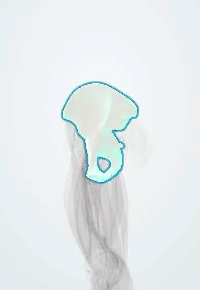
Hip bone !
The hip bone forms each side of the pelvis from three fused bones (ilium, ischium and pubis); its socket holds the ball of the femur.
Opens in the Durgical app, no sign-up needed.
What it is
The large bone on each side of the pelvis. It forms from three bones that fuse in the teenage years: the ilium (the wing you feel at your waist), the ischium (the sitting bone) and the pubis (at the front).
What it does
Joins the spine (through the sacrum) to the legs and carries the body's weight. Its cup-shaped socket, the acetabulum, holds the ball of the femur. Many hip and thigh muscles attach to it.
Key facts
- Joints with
- Sacrum (sacroiliac joint); opposite hip bone (pubic symphysis); femur (hip joint)
- Landmarks
- Ilium, ischium and pubis fused at acetabulum; iliac crest; ASIS and AIIS; ischial spine and tuberosity; pubic tubercle; obturator foramen; greater sciatic notch; lesser sciatic notch (obturator internus tendon, pudendal nerve, internal pudendal vessels); iliac tubercle; pecten pubis and pubic crest
Relations
- Above
- Iliac crest: attachments of the abdominal wall muscles, latissimus dorsi and quadratus lumborum
- In front
- Under the inguinal ligament, over the superior pubic ramus and the front of the ilium: iliopsoas, the femoral nerve, femoral artery and femoral vein; the lateral femoral cutaneous nerve passes close to the anterior superior iliac spine
- Medial
- Iliacus in the iliac fossa; obturator internus on the inner wall, with the obturator nerve and vessels leaving through the obturator canal
- Lateral
- Gluteal surface with gluteus maximus, gluteus medius and gluteus minimus; acetabulum with the femoral head
- Behind
- Greater sciatic notch: the superior gluteal vessels and nerve pass above piriformis; the sciatic nerve, inferior gluteal vessels and pudendal nerve pass below it, the pudendal nerve hooking round the ischial spine
- Below
- Ischial tuberosity with the hamstring origins; the ischioanal fossa lies medial to it
How to find it
The iliac crest is traced from the anterior superior iliac spine (ASIS) back to the posterior superior iliac spine (skin dimple, about S2). A line joining the highest points of the crests crosses the spine at about L4 (Tuffier's line), a lumbar puncture landmark. The pubic tubercle is felt at the lateral end of the pubic crest, and the ischial tuberosity deep in the buttock with the hip bent.
Examination
- Pelvic compression (springing): pressing the iliac wings together or apart causes pain in a pelvic ring injury; in an unstable patient it is done once at most, because it can move the fragments and worsen bleeding
- In a suspected apophyseal avulsion, point tenderness over the ASIS, the anterior inferior iliac spine or the ischial tuberosity, with pain on resisted contraction of the muscle attached there, locates the injury
On imaging
AP pelvis is the trauma screening view; inlet and outlet views and CT for ring fractures; Judet oblique views or CT for acetabular fractures. On the AP view the iliopectineal line marks the anterior column, the ilioischial line the posterior column and the teardrop the medial wall.
Common problems
- Fractures of the pelvis or socket, from high-energy injuries such as car accidents, or from falls in people with weak bones.
- Hip dysplasia: a shallow socket that covers too little of the ball, which can lead to labral tears and early arthritis.
- Hip impingement (FAI, pincer type): extra bone on the socket rim pinches the labrum.
- In teenagers, a strong muscle pull can break off a small piece of bone where the muscle attaches (avulsion).
How it is diagnosed
Physical exam; X-rays; CT scan for fractures and surgical planning; MRI for the labrum, cartilage and soft tissues.
Treatment
- Many stable fractures and avulsions heal with rest, protected weight-bearing and physical therapy.
- Surgery to fix unstable fractures with plates and screws.
- For dysplasia or impingement: physical therapy, or surgery to reshape or reorient the socket (for example hip arthroscopy or a periacetabular osteotomy).
Clinical notes
- Apophyseal avulsions in adolescent athletes: ASIS (sartorius), anterior inferior iliac spine (rectus femoris), ischial tuberosity (hamstrings)
- A pelvic binder centred on the greater trochanters closes an open-book injury and helps limit bleeding
- Iliac crest bone graft: anterior harvest can injure the lateral femoral cutaneous nerve; the posterior crest is also the usual site for bone marrow biopsy
- A shallow acetabular roof that covers the femoral head poorly underlies developmental dysplasia of the hip, screened in infants with the Ortolani and Barlow tests and hip ultrasound
Anatomical variants
- Sex differences: the female pelvis is wider and shallower, with a broader greater sciatic notch and a wider subpubic angle
- Caldwell–Moloy inlet shapes: gynecoid (the commonest in women), android, anthropoid and platypelloid (rare)
Development
Three primary centres (ilium, ischium, pubis) plus secondary centres at the triradiate cartilage, iliac crest, anterior inferior iliac spine and ischial tuberosity. The iliac crest apophysis ossifies from front to back, and its progress (Risser sign) is used to judge skeletal maturity.
Related conditions
- Femoral hernia
- Hip Osteoarthritis
- Developmental dysplasia of the hip
- Hip dislocation
- Meralgia paresthetica
- Sacroiliac joint pain
- Paget disease of bone
- Falls in older adults
- Acute leukaemia (AML and ALL)
- Febrile neutropenia
- Aplastic anaemia
- Myelodysplastic syndromes
- Paroxysmal nocturnal haemoglobinuria
- MGUS and Waldenström macroglobulinaemia
- Pelvic fracture
- Ewing sarcoma
Test yourself
Think of the answer, then tap the question to see it.
Which three bones form the hip bone, and where do they meet?
The ilium, ischium and pubis, which meet at the Y-shaped triradiate cartilage in the acetabulum and fuse in the mid-teens.
Why should a second injury be sought when the pelvic ring is fractured?
The ring rarely breaks in only one place: a second fracture or a joint disruption is usually present.
Which apophyseal avulsions of the hip bone occur in adolescent athletes, and which muscle pulls each?
ASIS (sartorius), anterior inferior iliac spine (rectus femoris) and ischial tuberosity (hamstrings).
What level does Tuffier's line, joining the highest points of the iliac crests, cross?
About L4; it is a landmark for lumbar puncture.
Educational information, not medical advice. For any symptom, see a health professional.
Its names
- Official Latin (TA2)
- os coxae TA2 1307
- Official Spanish (DeCS)
- Huesos pélvicos DeCS D010384
- English (TA2)
- Hip bone
- Also called
- coxal bone · Pelvic Bones · Ilium · pelvic bone · innominate bone · os pelvicum · Hip bones
Related
- Acetabular labrum
- Acetabular cartilage
- Sacroiliac ligaments
- Adductor longus
- Adductor longus tendon
- Adductor magnus
- Fascia lata
- Inferior gemellus
Sources: Terminologia Anatomica (TA2); DeCS; Anatomy, Bony Pelvis and Lower Limb: Pelvic Bones; Anatomy, Abdomen and Pelvis: Bones (Ilium, Ischium, and Pubis); Anatomía humana (4.ª ed.), tomo 1, cap. 63: Huesos del miembro inferior.
Spotted a mistake?We are always improving for the community. Tell us what is wrong and our team will review it.Report a mistake
Representative model, for educational purposes only.